Small molecule noncoding RNA gene hsa-mir-101 and antineoplastic use thereof
A tumor and tumor cell technology, applied in anti-tumor drugs, DNA/RNA fragments, recombinant DNA technology, etc., can solve the problems of unseen hsa-miR-101 and primary liver cancer research reports, etc., to reduce proliferation and incidence. Tumor ability, the effect of promoting sensitivity
- Summary
- Abstract
- Description
- Claims
- Application Information
AI Technical Summary
Problems solved by technology
Method used
Image
Examples
Embodiment 1
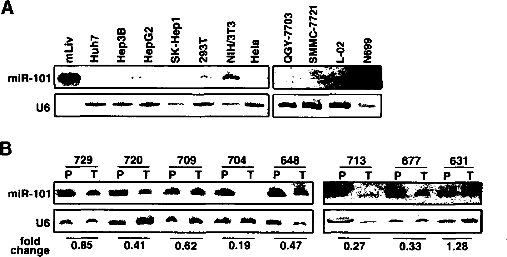
[0014] Example 1: Detection of the expression level of hsa-miR-101 in liver cancer tissue specimens
[0015] Extract total RNA from liver cancer and non-cancerous liver tissues, mix the RNA sample with RNA loading buffer in a ratio of 3:1, denature at 80°C for 10 minutes, and then place it on ice for 5 minutes; Under 10W electric power, use 15% denaturing polyacrylamide gel for about 40min electrophoresis separation; after electrophoresis is completed, cut the corners of the gel and mark, assemble the gel and membrane in order in the semi-dry transfer tank, constant flow 8mA / cm 2 Transfer the membrane for 1 hour; remove the Hybond N+ nylon membrane and cut the corner to make a mark, 70,000μJ / cm 2 UV cross-link and dry bake at 80°C for 2h; after wetting the nylon membrane with 2×SSC, spread it on the inner wall of the hybridization tube, and remove the air bubbles between the membrane and the tube wall, add the pre-hybridization solution, and put it into the hybridization box. Rota...
Embodiment 2
[0017] Example 2: Overexpression of hsa-miR-101
[0018] Introducing the small RNA of hsa-miR-101 into cells is achieved by using Invitrogen's transfection reagent Lipofectamine RNAi MAX to reverse. Take a 24-well plate as an example. Other well plates can be scaled up according to the size of the wells. Or shrink: trypsinize the cells and resuspend them in DMEM medium containing 10% FBS without dual antibody to make the cell density 3×10 4 Cells / ml; dilute small RNA in 100μl opti-MEM, tap the tube wall to mix; add 1μl Lipofectamine RNAi MAX to each tube, tap the tube wall to mix, and leave it at room temperature for 15 minutes; add RNAiMAX / siRNA diluent Then add 500μl of cell diluent to the 24-well plate (to make the cell density approximately 30-50% confluent after 24h of transfection), mix well and incubate in an incubator for a certain period of time before performing related functional tests.
Embodiment 3
[0019] Example 3: Detection of cell apoptosis
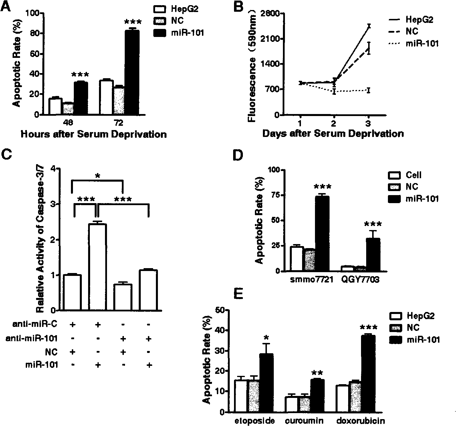
[0020] After processing the cells as needed, aspirate the culture supernatant, fix with 4% paraformaldehyde at room temperature for 15 minutes, stain with 1μg / ml (final concentration) DAPI staining solution at room temperature for 5 minutes, and observe the nucleus morphology under a Lycra fluorescence microscope. Cells with chromatin constriction and DNA fragmentation are considered to be apoptotic cells, and the apoptosis rate is calculated according to the following method:
[0021] Apoptosis rate (%) = number of apoptotic cells / total number of cells (total number of cells ≥500)
[0022] We tested the effect of hsa-miR-101 on tumor cell apoptosis under the conditions of serum starvation and chemotherapeutic drug treatment, and found that the experimental group overexpressing hsa-miR-101 in HepG2 cells (ATCC in the United States) died after 48 hours of serum starvation. The death rate is ~2 times that of the negative control (NC) g...
PUM
 Abstract
Description
Claims
Application Information
 - R&D
- Intellectual Property
- Life Sciences
- Materials
- Tech Scout
- Unparalleled Data Quality
- Higher Quality Content
- 60% Fewer Hallucinations
Browse by: Latest US Patents, China's latest patents, Technical Efficacy Thesaurus, Application Domain, Technology Topic, Popular Technical Reports.
© 2025 PatSnap. All rights reserved.Legal|Privacy policy|Modern Slavery Act Transparency Statement|Sitemap|About US| Contact US: help@patsnap.com


