Real time fluorescent PCR method for detecting aquatic product food allergen gene

A technology of food allergy and real-time fluorescence, which is applied in biochemical equipment and methods, fluorescence/phosphorescence, microbial measurement/inspection, etc., to achieve the effect of high sensitivity and short detection time

Inactive Publication Date: 2009-05-20
JIMEI UNIV
View PDF0 Cites 10 Cited by
  • Summary
  • Abstract
  • Description
  • Claims
  • Application Information

AI Technical Summary

Problems solved by technology

However, there is no real-time fluorescent PCR method to detect aquatic food allergen genes. Ther

Method used


Image

  • Real time fluorescent PCR method for detecting aquatic product food allergen gene
  • Real time fluorescent PCR method for detecting aquatic product food allergen gene
  • Real time fluorescent PCR method for detecting aquatic product food allergen gene

Examples


Embodiment 1

[0030] Embodiment 1: be used for detecting the real-time fluorescent PCR of carp Par gene

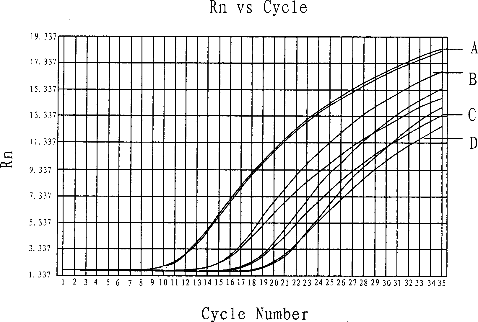
[0031] The Par gene was detected on carp muscle total RNA diluted 10 times by using TaqMan probe fluorescent PCR method. Par gene upstream primer 0.5 μL, Par gene downstream primer 0.5 μL, Par gene probe 0.3 μL, Oligo dT 15 1 μL and 0.4 μL of RNA template. The reaction parameters were reverse transcription at 48°C for 30 minutes; pre-denaturation at 94°C for 10 minutes; 94°C / 40S, 55°C / 45S, 72°C / 90S, a total of 35 cycles. The results showed that the amplification kinetic curve was a typical S-type, and gradually shifted backwards with the decrease of sample concentration; the Ct value gradually increased with the decrease of sample concentration (such as figure 1 shown).

[0032] exist figure 1 In the middle, the kinetic curve of TaqMan probe fluorescent PCR (Par gene) amplification of 10-fold serially diluted carp RNA; A.1:10 dilution, Ct=15.3089; B.1:100 dilution, Ct=16.5373; C.1 :...

Embodiment 2

[0033] Example 2: Real-time fluorescent PCR for detection of Scylla serrata TM gene

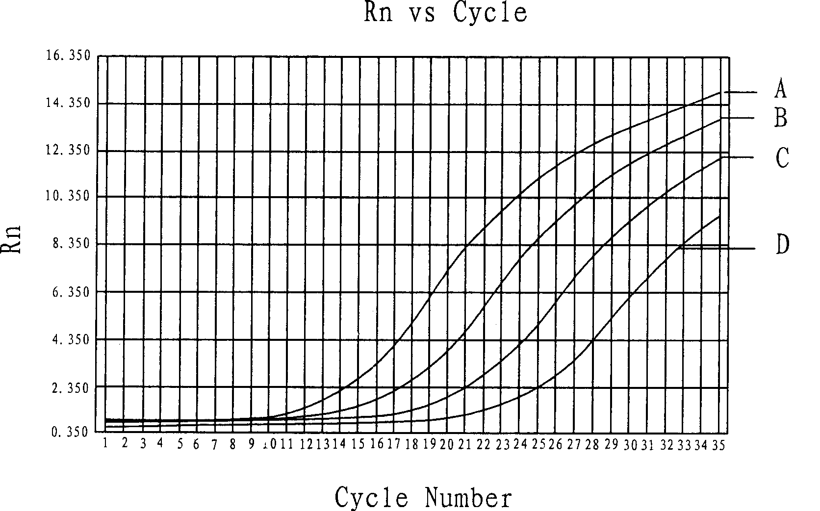
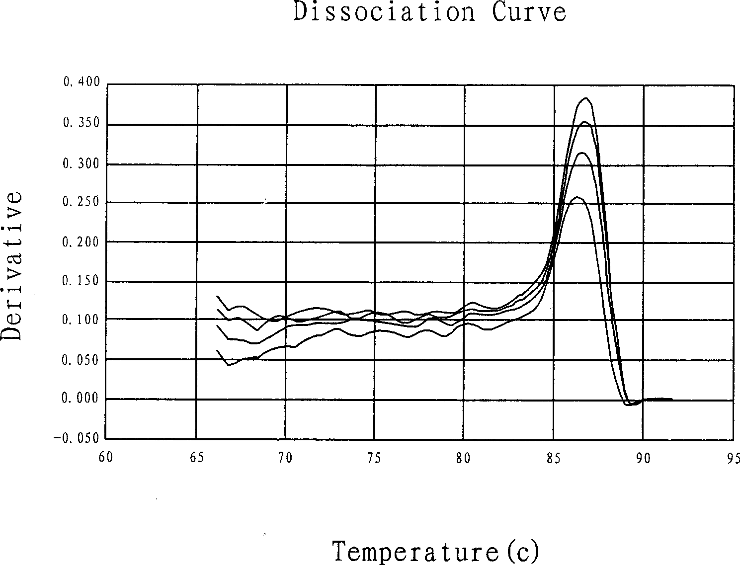
[0034] The TM gene was detected on the 10-fold serially diluted muscle total RNA of Scylla serrata by SYBR Green fluorescent PCR method. The reaction system was PCR Master Mix 10 μL, sterilized ultrapure water 6.7 μL, MultiScribe Reverse Transcriptase 0.5 μL, RNase Inhibiter 0.4 μL, TM gene upstream primer 0.5 μL, TM gene downstream primer 0.5 μL, Oligo dT 15 1 μL and 0.4 μL of RNA template. The reaction parameters are reverse transcription at 48°C for 30 minutes; pre-denaturation at 94°C for 10 minutes; 94°C / 40S, 55°C / 45S, 72°C / 90S, a total of 35 cycles; set the melting curve analysis from 60°C to 95°C. The results showed that the amplification kinetic curve was a typical S-type, and gradually shifted backwards with the decrease of sample concentration; the Ct value gradually increased with the decrease of concentration (such as Figure 2A shown); the melting curve profile of the amplifie...

Embodiment 3

[0036] Example 3: Fish Par gene detection in unknown samples

[0037] The detection of the Par gene in the fish Par gene in the unknown sample was carried out by using the TaqMan probe fluorescent PCR method, and the specific conditions refer to Example 1. The results showed that the amplification kinetic curve of the unknown sample was a typical S-type, and no change in the fluorescent signal was detected in the negative control, indicating that the unknown sample contained the fish Par gene (such as image 3 shown).

[0038] exist image 3 In, the kinetic curve of TaqMan probe fluorescent PCR detection of fish Par gene in unknown samples; A. Sample S-I, Ct=15.2695; B. Sample S-II, Ct=16.0500; C. Negative control, Ct=Undet.


PUM


Abstract

The invention discloses a real-time fluorescent PCR method for testing the allergen genes of aquatic food, comprising the following steps: total RNA of samples is extracted; the allergen genes Par and TM of aquatic food, and internal standard gene Beta-actin are respectively tested by SYBR Green or TaqMan probe real-time fluorescent PCR technology; and result analysis is carried out to the tested samples according to the amplification kinetic curve and the Ct value. As the real-time fluorescent PCR technology is used for testing the allergen genes Par and TM of aquatic food, compared with other methods, the method has the advantages of high sensitivity, short testing time, available quantitative analysis, and the like, and can be used in the qualitative screening, quantitative analysis and confirmation identification of allergen genes in unknown species or mixed samples of aquatic food.

Description

technical field [0001] The invention relates to the detection of food allergen genes, in particular to a real-time fluorescent polymerase chain reaction (PCR) method for detecting aquatic product food allergen genes, which is mainly used for fish parvalbumin (Par) and Real-time fluorescent PCR detection of the tropomyosin (TM) gene in crustaceans. Background technique [0002] Food allergy is also called allergic reaction in medicine. Substances that cause food allergic reactions are called food allergens, such as fish, shrimp, crab, milk, eggs, etc. The reaction mechanism is that after the allergen enters the body, it will cause the body to have a normal or excessive immune response, that is, a hypersensitivity reaction. When exposed to the same allergen again, multiple IgE molecules bind to the antibody, causing changes in the structure of the Fc end of IgE, leading to cell degranulation, releasing histamine, serotonin, leukotrienes and other biological respiratory activ...

Claims


Application Information

Patent Timeline
IPC IPC(8): C12Q1/68G01N21/64
Inventor 刘光明曹敏杰蔡慧农
Owner JIMEI UNIV
Why Eureka
Try Eureka