Method for constructing and implanting organization engineering liver implant
A construction method and tissue engineering technology, applied in medical science, prosthesis, etc., can solve problems such as difficulty in providing functional support, rapid inactivation, and inability to induce liver parenchyma, so as to promote effective contact, improve implantation, and facilitate diffusion and the effect of nutrient transport
- Summary
- Abstract
- Description
- Claims
- Application Information
AI Technical Summary
Problems solved by technology
Method used
Image
Examples
Embodiment 1
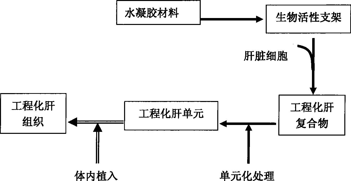
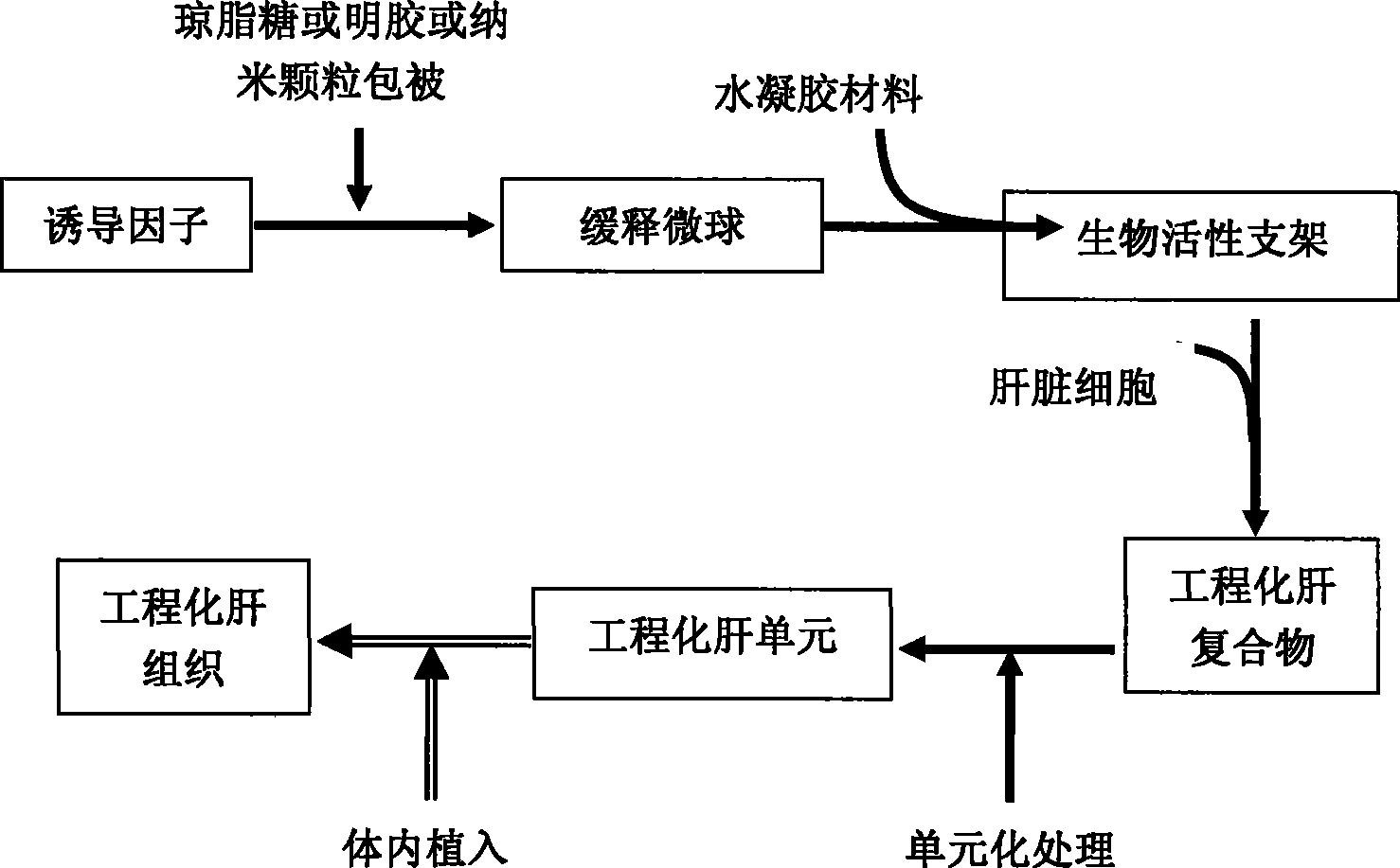
[0022] Example 1 Construction of engineered liver graft unit
[0023] Dissolve the growth factors in 5% agarose, the final concentrations of the growth factors HGF and VEGF are 5 μg / ml respectively, after the agarose coagulates, mechanically break it into small microspheres to obtain agarose Slow-release microspheres.
[0024] Then operate on ice, compound the gel sustained-release microspheres and hydrogel materials to construct a bioactive scaffold; mix liquid type I collagen (from SD rat tail tendon) and 2×DMEM (Sigma, US) in equal proportions, Adjust the pH to 7.4 with 0.1mol / L NaOH solution to obtain a mixture, add gel slow-release microspheres to construct a bioactive scaffold.
[0025] Primary isolated liver cells (SD rats, 200-250 g) were mixed with bioactive scaffolds (on ice) to create liver cell / scaffold complexes. Aspirate the liver cell / scaffold complex into a 1 ml disposable syringe and place at 37 °C, 5% CO 2 Incubate in an incubator for 15 minutes, push out ...
Embodiment 2
[0027] The prepared liver unit was subcutaneously implanted to construct engineered liver tissue. The experiment was carried out as follows:
[0028] Liquid type I collagen (from the tail tendon of SD rats) and 2×DMEM medium were mixed in equal proportions, and the pH value of the mixture was adjusted to 7.2-7.4 with 0.1 mol / L NaOH. Freshly isolated hepatocytes were combined with the above mixture to construct hepatocyte / collagen gel complexes, and placed at 37°C, 5% CO 2 Incubate for 10 minutes in the incubator. After the complex forms a gel, it is broken up into small pieces of hepatic cell units. After creating a subcutaneous pouch in the armpit of the rat, hepatocyte units were implanted into the pouch.
[0029] The results of in vitro experiments show that after the hepatocytes are compounded with liquid type I collagen, the cells are evenly distributed in the whole complex and can grow in three dimensions; during the whole in vitro culture process, the hepatocytes alw...
Embodiment 3
[0031] Construction of engineered liver tissue in situ in the liver using bone marrow stromal stem cells as seed cells
[0032]Bone marrow stromal stem cells of SD rats cultured to the fourth generation by conventional methods were compounded with liquid type I collagen, concentrated DMEM (2×) culture medium and commercially available hepatocyte culture medium, and growth factors HGF and VEGF were added, and the final concentrations were respectively 5 μg / ml. Aspirate the complex into a 1ml syringe and place it in a 37°C, 5% CO2 incubator for 15 minutes, push it out after the complex forms a semi-solid gel, and cut it into small pieces with a length of 1mm to form A cylindrical liver graft unit with a diameter of 0.5 mm and a height of 1 mm.
[0033] The constructed liver graft unit was implanted into the liver of syngeneic rats by injection. One week later, liver-like tissue was formed in the transplanted area. Histological and immunohistochemical tests can be seen; 1) isl...
PUM
| Property | Measurement | Unit |
|---|---|---|
| Diameter | aaaaa | aaaaa |
| Height | aaaaa | aaaaa |
Abstract
Description
Claims
Application Information
 - R&D
- Intellectual Property
- Life Sciences
- Materials
- Tech Scout
- Unparalleled Data Quality
- Higher Quality Content
- 60% Fewer Hallucinations
Browse by: Latest US Patents, China's latest patents, Technical Efficacy Thesaurus, Application Domain, Technology Topic, Popular Technical Reports.
© 2025 PatSnap. All rights reserved.Legal|Privacy policy|Modern Slavery Act Transparency Statement|Sitemap|About US| Contact US: help@patsnap.com


