Orbital margin tissue engineering bone and application thereof

A technology of tissue engineering bone and tissue engineering, applied in bone implants, medical science, prostheses, etc., to achieve the effect of saving experimental time, convenient operation, and non-immunogenicity

Inactive Publication Date: 2010-01-20
SHANGHAI NINTH PEOPLES HOSPITAL AFFILIATED TO SHANGHAI JIAO TONG UNIV SCHOOL OF MEDICINE
View PDF3 Cites 5 Cited by
  • Summary
  • Abstract
  • Description
  • Claims
  • Application Information

AI Technical Summary

Problems solved by technology

However, there is no report about a kind of orbit

Method used


Image

  • Orbital margin tissue engineering bone and application thereof
  • Orbital margin tissue engineering bone and application thereof
  • Orbital margin tissue engineering bone and application thereof

Examples


Embodiment 1

[0044] Construction of canine orbital rim tissue-engineered bone in vitro

[0045] 1. BMSCs in vitro osteogenic induction culture and detection

[0046] BMSCs were cultured in α-MEM culture medium supplemented with β-glycerol phosphate, dexamethasone and ascorbic acid for in vitro osteogenic induction, and uninduced BMSCs were used as controls to detect cell morphology and osteogenic activity. The results showed that the cell growth cycle and cell proliferation of canine BMSCs were not affected after osteogenic induction culture, and they could be passed on for more than 10 generations. Calcium nodules stained strongly positive; express osteogenic marker proteins (OCN, OPN, BSP, Cbfa1, type I collagen, etc.) and osteogenic related genes (OCN, OPN, BSP). The ALP staining of the uninduced BMSCs was weakly positive, the calcium nodule staining was negative, and the expression levels of osteogenic marker proteins and related genes were lower than those of the induced group.

[0...

Embodiment 2

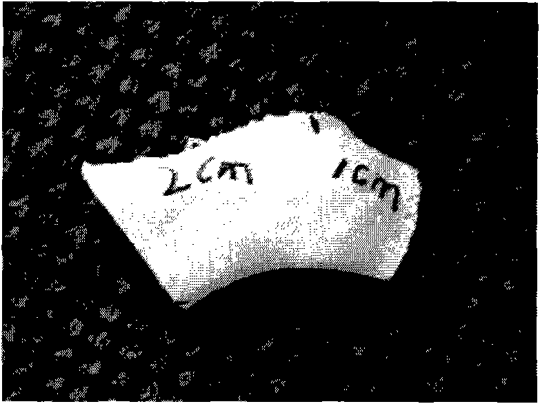
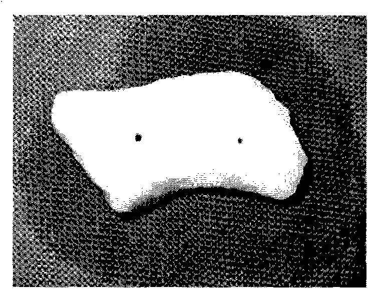
[0052] The Experiment of Heterotopic Osteogenic Ability of Orbital Marginal Tissue-Engineered Bone Constructed in Vitro

[0053] According to the method of Example 1, autologous induced BMSCs and β-TCP scaffold materials were combined in vitro to construct tissue engineered bone.

[0054] Three pieces of materials were subcutaneously implanted on the back of each dog: one piece of autologous induced BMSCs combined with TCP, one piece of autologous uninduced BMSCs combined with TCP, and one piece of simple TCP material. Surgical method: After the experimental animal dog was anesthetized, a small incision of 1.5 cm was made in three separate areas on the back of the dog, the surrounding subcutaneous tissue was fully freed, and the material was implanted, and the incision was sutured intermittently with No. 1 suture.

[0055] The specimens were taken out at 1 month, 2 months, and 3 months after the material implantation, and 9 pieces of subcutaneous specimens were taken from the ...

Embodiment 3

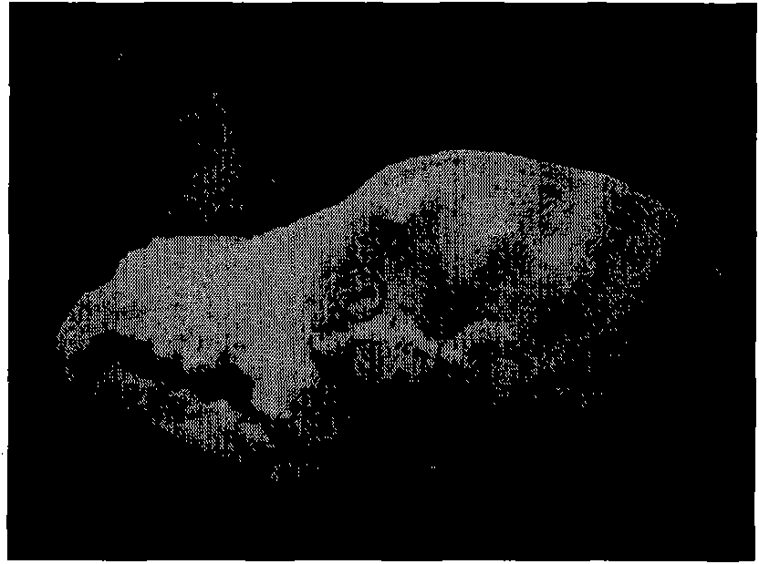
[0058] Experiment of Orbital Margin Tissue Engineering Bone in Situ Repairing Canine Orbital Margin Bone Defect

[0059] According to the method of Example 1, autologous induced BMSCs and β-TCP scaffold materials were combined in vitro to construct tissue engineered bone.

[0060] Establishment of bone defect model of canine infraorbital rim: skin incision was made on the lower eyelid, subcutaneous tissue and muscle were separated suddenly, the infraorbital rim and zygomatic arch were fully exposed, and the periosteum was incised and peeled off with a dissector. According to the location and size of the defect in the computer-aided design, a segmental bone defect with a length of about 25 mm was made from the medial infraorbital rim to the zygomatic arch using a chainsaw, and the periosteum at the defect was removed. Orbital rim tissue-engineered bone constructed by autologous induced BMSCs and β-TCP was implanted, and a small hole was drilled with an electric drill at the two...


PUM

PropertyMeasurementUnit
Inoculation densityaaaaaaaaaa

Abstract

The invention relates to an orbital margin tissue engineering bone and application thereof. The orbital margin tissue engineering bone is prepared by the following steps: 1, culturing and inducing bone seed cells; 2, preparing an individual bone repair scaffold; and 3, combining the bone seed cells and the bone repair scaffold in vitro and culturing, wherein the bone seed cells cultured to a second generation are combined with the bone repair scaffold in the step 3 to form a compound of the bone seed cells and the bone repair scaffold. Similar to the tissue structures and characteristics of normal orbital margins, the orbital margin tissue engineering bone constructed and cultured in vitro can facilitate autogenous bone regeneration while a bracket material is degraded, generate rational bone reconstruction and finally be replaced by autogenous bone tissues in vivo so as to realize dual-repair of the shape and the function.

Description

technical field [0001] The invention relates to a tissue engineered bone, in particular to an orbital edge tissue engineered bone and its application. Background technique [0002] The orbital rim is an irregular circular opening. As the most central part of the face, it not only participates in the shaping of the facial contour, but also plays an important role in protecting the eyeballs. Congenital malformations, tumor surgery, trauma, and inflammation may cause bone defects or hypoplasia of the orbital rim, and it is necessary to repair and reconstruct the fractured, defective, and deformed orbital rims. At present, the orbital rim repair materials used at home and abroad include autologous bone, allogeneic bone and various artificial materials. [0003] Autologous bone transplantation has always been the gold standard for various clinical bone repair operations, but the source of autologous bone is limited, and there are risks of complications such as bleeding, infectio...

Claims


Application Information

Patent Timeline
IPC IPC(8): A61L27/36A61L27/12A61F2/28
Inventor 范先群周慧芳
Owner SHANGHAI NINTH PEOPLES HOSPITAL AFFILIATED TO SHANGHAI JIAO TONG UNIV SCHOOL OF MEDICINE
Why Eureka
Try Eureka