Method for screening and identifying adhesion proteins

An adhesion protein and protein technology, which is applied in the field of screening and identification of proteins, can solve the problems of high false positive rate of adhesion proteins, no adhesion function, complex intestinal environment, etc., and achieves the effect of low cost and improved efficiency.

Inactive Publication Date: 2010-11-10
NANCHANG UNIV
View PDF2 Cites 6 Cited by
  • Summary
  • Abstract
  • Description
  • Claims
  • Application Information

AI Technical Summary

Problems solved by technology

However, due to the fact that the characteristic sequence of the adhesion protein has not yet been confirmed, coupled with the complexity of the intestinal environment, a considerable part of the inferred protein does not have the adhesion function, so this strategy has a certain degree of blindness
[0004] This method combines protein technology, cell culture and molecular immune meth

Method used


Image

  • Method for screening and identifying adhesion proteins
  • Method for screening and identifying adhesion proteins
  • Method for screening and identifying adhesion proteins

Examples


Embodiment 1

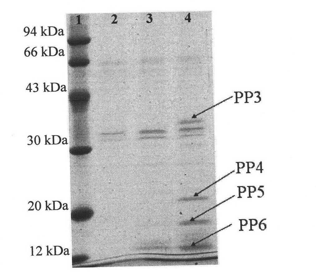
[0016] Example 1: Screening of Bifidobacterium longum adhesion proteins by magnetic bead method

[0017] 1. Activation of magnetic beads: Wash 30 mg of carboxylated magnetic beads with 6 mL of sodium phosphate buffer (0.2 M, pH 4.5) for 3 times, resuspend in 1.5 mL of sodium phosphate buffer, add 2.25 mg of carbodiimide and 1.5 mg thiohydroxysuccinimide, react at room temperature for 40min.

[0018] 2. Adjust the pH of the above reaction system to 7.8, add 1500 mg of intestinal epithelial cell HT-29 cell fragments, react at room temperature for 3 hours, and overnight at 4°C.

[0019] 3. Adsorb the magnetic beads on the magnetic stand to remove the solution, wash the magnetic beads 3 times with PBS to wash away the unbound intestinal epithelial cell fragments, add 1.5mL 3% BSA solution to seal the magnetic beads for 1h, adsorb the magnetic beads on the magnetic stand to remove the solution, wash with PBS Wash the magnetic beads 3 times.

[0020] 4. Divide the magnetic beads i...

Embodiment 2

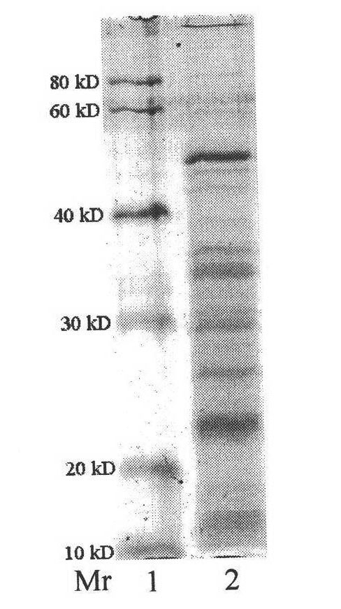
[0023] Example 2: Screening of Adhesive Proteins of Bifidobacterium adolescentis by Magnetic Beads Method

[0024] 1. Activation of magnetic beads: wash 10 mg of carboxylated magnetic beads with 2 mL of sodium phosphate buffer (0.2 M, pH 4.5) for 3 times, resuspend in 0.5 mL of sodium phosphate buffer, add 0.75 mg of carbodiimide and 0.5 mg thiohydroxysuccinimide, react at room temperature for 40min.

[0025] 2. Adjust the pH of the above reaction system to 7.8, add 500 mg of intestinal epithelial cell HT-29 cell fragments, react at room temperature for 3 hours, and overnight at 4°C.

[0026] 3. Adsorb the magnetic beads on the magnetic stand to remove the solution, wash the magnetic beads 3 times with PBS to wash away the unbound intestinal epithelial cell fragments, add 1.5mL 3% BSA solution to seal the magnetic beads for 1h, adsorb the magnetic beads on the magnetic stand to remove the solution, wash with PBS Wash the magnetic beads 3 times.

[0027] 4. Add 0.2mL 2.5mg / mL...

Embodiment 3

[0030] Example 3: Magnetic bead method for screening Lactobacillus plantarum adhesion proteins

[0031] 1. Activation of magnetic beads: wash 10 mg of carboxylated magnetic beads with 2 mL of sodium phosphate buffer (0.2 M, pH 4.5) for 3 times, resuspend in 0.5 mL of sodium phosphate buffer, add 0.75 mg of carbodiimide and 0.5 mg thiohydroxysuccinimide, react at room temperature for 40min.

[0032] 2. Adjust the pH of the above reaction system to 7.8, add 500 mg of intestinal epithelial cell HT-29 cell fragments, react at room temperature for 3 hours, and overnight at 4°C.

[0033] 3. Adsorb the magnetic beads on the magnetic stand to remove the solution, wash the magnetic beads 3 times with PBS to wash away the unbound intestinal epithelial cell fragments, add 1.5mL 3% BSA solution to seal the magnetic beads for 1h, adsorb the magnetic beads on the magnetic stand to remove the solution, wash with PBS Wash the magnetic beads 3 times.

[0034] 4. Add 0.2 mL of Lactobacillus pla...


PUM


Abstract

The invention provides a method for screening and identifying adhesion proteins. The method is characterized by comprising the following steps of: carboxylating superficial magnetic beads; processing the carboxylated magnetic beads by using carbodiimide and sulfo-hydroxysuccinimide; covalently coupling the magnetic beads with enterocyte debris to form amido bonds; after cleaning the magnetic beads, incubating the magnetic beads and probiotic surface protein solution together for one hour; washing the magnetic beads by using PBS buffer solution for three times to remove non-adhesion surface proteins; eluting the adhesion proteins by using 50 mM of glycine hydrochloric acid buffer solution of which the pH value is 2.3; and after SDS-PAGE electrophoresis, sequencing by mass spectrometry to identify the adhesion proteins. The method for screening and identifying the adhesion proteins has the advantages that: a magnetic bead method makes use of the characteristic that the adhesion proteins can adhere enterocyte to combine the function and separation into one step, so that the efficiency of screening the adhesion proteins is greatly improved, and the adhesion proteins can be efficiently screened and identified in a high-throughput mode; and compared with the conventional chromatography method, the method has the advantages of low cost, time saving, labor saving, and capacity of screening and identifying a plurality of adhesion proteins in one step.

Description

technical field [0001] The invention relates to a method for screening and identifying proteins, in particular to a method for screening and identifying adhesion proteins. Background technique [0002] The study of the interaction between microorganisms and the environment (host) is the core research direction in the field of microbiology today. In recent years, the colonization and reproduction mechanism of intestinal flora in the intestinal tract has attracted widespread attention, especially its special genetic characteristics, secretion of proteins and biologically active substances with potential biological significance, and its adaptability in the intestinal environment. , and the interaction mechanism with host cells (intestinal epithelial cells) is intricate. There are still scientific research blind spots in the transmission of substances / signals, changes in the internal and external environment, and the infection of pathogenic bacteria. The molecular mechanism of ...

Claims


Application Information

Patent Timeline
IPC IPC(8): G01N27/62G01N27/447
Inventor 魏华徐锋彭珍谭强来徐迪陈廷涛熊勇华
Owner NANCHANG UNIV
Why Eureka
Try Eureka