
Detection method of ATP (Adenosine Triphosphate) content and ATP aptamer sensor

An aptamer sensor and detection method technology, which is applied to biochemical equipment and methods, microbial determination/inspection, instruments, etc., can solve problems such as high cost and complicated operation, and achieve the effect of simple method

Active Publication Date: 2011-01-05
CHANGZHOU INST OF ENERGY STORAGE MATERIALS &DEVICES
View PDF3 Cites 13 Cited by
  • Summary
  • Abstract
  • Description
  • Claims
  • Application Information

AI Technical Summary

Problems solved by technology

In the prior art, in the detection process of ATP, the nucleic acid aptamer needs to be chemically labeled, and then detected according to the changes of the label itself or its catalytic substrate before and after the recognition reaction, so the operation is complicated and the cost is high

Method used


Image

  • Detection method of ATP (Adenosine Triphosphate) content and ATP aptamer sensor
  • Detection method of ATP (Adenosine Triphosphate) content and ATP aptamer sensor

Examples


Embodiment 1

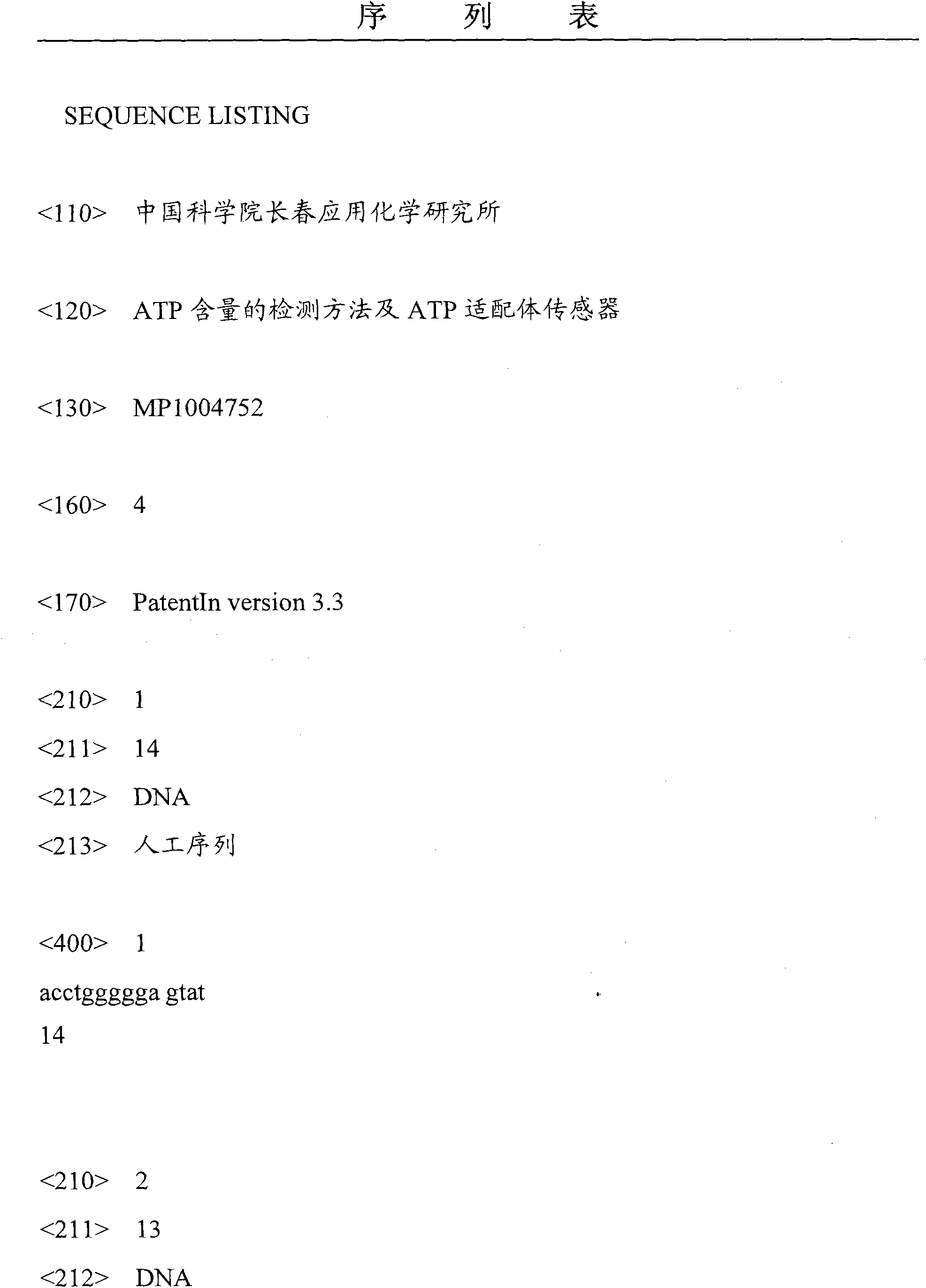
[0047] Divide the chain containing the ATP aptamer into two sequences, as shown in SEQ ID No.1 and SEQ ID No.2, which are the 3'end of the first strand in the single-stranded DNA and part of the double-stranded DNA;

[0048] The second strand in the partial DNA double strand is provided, as shown in SEQ ID No. 3, the second strand in the DNA double strand is complementary to the 5'end of the first strand in the partial DNA double strand, forming a partial DNA double strand (part ds -DNA).

Embodiment 2

[0050] After modifying the sulfhydryl group at the 5'end of the DNA single strand, the sequence 5'HS-(CH 2 ) 6 -ACCTGGGGGAGTAT-3’;

[0051] Dip the gold electrode (diameter 3mm) into the 5μM DNA single-stranded solution prepared in Example 1, and take it out after 1 hour;

[0052] After washing, the gold electrode is placed in a 10 mM mercaptohexanol (MCH) solution for 30 minutes;

[0053] Immerse the gold electrode placed in the mercaptohexanol (MCH) solution in the mixed solution of 5μM part ds-DNA and 5μM ATP prepared in Example 1, and take it out after reacting for 30 minutes;

[0054] Finally, put the gold electrode in 20mM Ru(phen) 3 2+ Reaction in solution for 1h.

[0055] ECL detection was carried out in 0.2M phosphate buffered saline (PBS) (pH 7.5, containing 20mM oxalate as a co-reactant), and cyclic voltammetry scanning was carried out in the range of 0-1.3 volts with a sweep rate of 0.05 volts / second.

Embodiment 3

[0057] After modifying the sulfhydryl group at the 5'end of the DNA single strand, immerse the gold electrode (3mm in diameter) in the 5μM DNA single strand solution prepared in Example 1, and take it out after 1 hour;

[0058] After washing, the gold electrode is placed in a 10 mM mercaptohexanol (MCH) solution for 30 minutes;

[0059] Dip the gold electrode placed in the mercaptohexanol (MCH) solution into the mixed solution of 5μM part ds-DNA and 10μM ATP prepared in Example 1, and take it out after reacting for 30 minutes;

[0060] Finally, put the gold electrode in 20mM Ru(phen) 3 2+ React in solution for 1h.

[0061] The ECL detection was performed in 0.2M PBS (pH 7.5, containing 20mM oxalate as a co-reactant), and the cyclic voltammetry scan was performed in the range of 0-1.3 volts, and the scanning rate was 0.05 volts / sec.


PUM


Abstract

The embodiment of the invention discloses a detection method of ATP (Adenosine Triphosphate) content and an ATP aptamer sensor. The detection method of the ATP content comprises the following steps of: providing a gold electrode the surface of which is fixed with a DNA single strand, wherein the DNA single-stranded sequence is shown as SEQ ID No.1; providing part of DNA double strands, wherein the tail end of the first strand 3' in the partial DNA double strands is shown as SEQ ID No.2 and the tail end of the strand 5' is complementary to the second strand; immersing the gold electrode in the mixed liquor of the partial DNA double strands and a sample to be tested; by using Ru(phen)3<2+>, [Ru(bpy)2dppz]<2+> or [Ru(phen)2(dppz)]<2+> as an electrochemical luminescence probe, performing ECL (Electrochemiluminescence) detection on the gold electrode. By using aromatic ring with larger ruthenium compound ligand, the invention has the performance of embedding the DNA double-stranded structure and realizes detection of the ATP; and besides, the detection method does not need to use chemical marks and is simple.

Description

Technical field [0001] The invention relates to the field of biotechnology, and more specifically, to a method for detecting ATP content and an ATP aptamer sensor. Background technique [0002] Adenosine triphosphate (ATP) exists in the cells of organisms ranging from microorganisms to higher animals and plants. Its main role is to provide energy and participate in the metabolism of fat, protein, sugar and nucleic acid in the body. It is an important source of energy for the body, and it maintains the normality of the organism. The function has an irreplaceable role. The rapid and efficient determination of ATP is of great significance for studying the physiological activity and metabolic process of cells and even the body, conducting drug sensitivity experiments, and food hygiene monitoring. [0003] Traditionally, ATP detection methods include electrophoresis, high performance liquid chromatography and isotope tracing methods. In the electrophoresis method, the sample needs to ...

Claims


Application Information

Patent Timeline
IPC IPC(8): G01N27/48G01N27/327C12Q1/68
Inventor 徐国宝刘中原
Owner CHANGZHOU INST OF ENERGY STORAGE MATERIALS &DEVICES
Learn More