Fluorescence in situ hybridization (FISH) method for fish chromosomes

A fluorescence in situ hybridization, chromosome technology, applied in fluorescence/phosphorescence, biochemical equipment and methods, microbial determination/inspection, etc. clear image effect

Inactive Publication Date: 2011-02-16
DALIAN OCEAN UNIV
View PDF0 Cites 7 Cited by
  • Summary
  • Abstract
  • Description
  • Claims
  • Application Information

AI Technical Summary

Problems solved by technology

FISH technology has been successfully used in humans and mammals, but its application in fish genetics research is not deep enough, and there are few application reports. For example, Ji Fuyun et al. (2003) used FISH technology to prove the existence of Hsl gene in eel ; Yi Meisheng et al. (2002) used FISH technology to compare the heterochromatin region of the long arm of the human Y chromosome with the fish genome
So far, there is no report on the application of FISH technology in fish polyploidy

Method used


Image

  • Fluorescence in situ hybridization (FISH) method for fish chromosomes
  • Fluorescence in situ hybridization (FISH) method for fish chromosomes
  • Fluorescence in situ hybridization (FISH) method for fish chromosomes

Examples


Embodiment 1

[0021] a. Preparation of chromosome slide specimens

[0022] Use methods such as erythrocyte nuclear volume measurement or flow cytometry to detect the ploidy of the collected loach, and screen the natural diploid. Chromosomal slide specimens were observed under a phase-contrast microscope, and metaphase specimens with clear images and well-dispersed chromosomes were selected and stored in a -80°C refrigerator.

[0023] b. Pretreatment of chromosome slide specimens

[0024] 1) Take out the chromosome slide specimens stored at -80°C and put them in a 65°C incubator for 2 hours to dry;

[0025]2) Soak the dried chromosome slide specimen in 4×SSC solution for 5 minutes, pipette 100 μL of 0.5 mg / ml RNase solution onto the chromosome slide specimen, cover the slide with parafilm, and place The slides were placed in a wet box with 2×SSC solution at the bottom at 37°C for 30min. Operate in groups of two slides;

[0026] 3) Carefully remove the parafilm with fine tweezers, immedia...

Embodiment 2

[0057] The difference from Example 1 is the preparation of chromosome slide specimens: use methods such as erythrocyte nuclear volume measurement or flow cytometry to detect the ploidy of the collected loach, and screen for natural tetraploids. According to the methods of the prior art, use Live gill tissue was used as material, and natural tetraploid chromosome slide specimens were prepared by air-drying method, observed under a phase-contrast microscope, and metaphase specimens with clear images and well-dispersed chromosomes were selected and stored in a -80°C refrigerator.

[0058] Other steps and signal observation are the same as in Example 1.

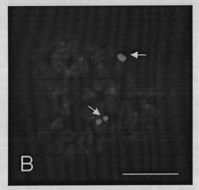
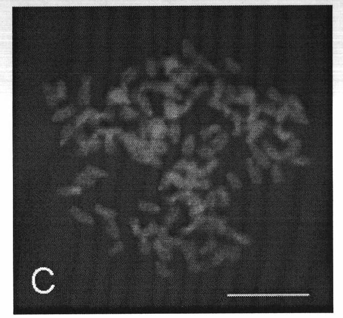
[0059] Metaphase (DAPI counterstaining) effect diagram and fluorescence in situ hybridization effect diagram of metaphase phase are as follows image 3 (C), Figure 4 (D) shown. Chromosomes were counterstained with DAPI, showing blue; arrows indicate hybridization signal sites (green); the scale bar is 10 μm.


PUM


Abstract

The invention discloses a fluorescence in situ hybridization (FISH) method for fish chromosomes. In the method, on the basis that a metaphase split phase specimen with a clear image and good chromosome spreading is obtained, a probe is marked by FISH technology and a nick translation method by taking Biotin-16-dUTP as a marker, a hybridization signal is amplified at two stages and a human 5.8S+28SrDNA probe is clearly positioned on a polyploid fish chromosome nucleolus organizer region (NOR). The chromosome ploidy and a karyotype of a polyploid are compared and analyzed by observing the chromosomal localization of ribosome 5.8S+28SrDNA on a polyploid fish, namely, the polyploid fish is proved to be a genetic polyploidy or an evolution polyploidy and an autopolyploid or an allopolyploid byanalyzing.

Description

Technical field: [0001] The invention relates to a fluorescent in situ hybridization method for fish chromosomes, in particular to a method with high sensitivity, strong specificity and accurate positioning, which is beneficial for analyzing whether polyploid fish are genetic polyploids or evolutionary polyploids, and whether they are polyploid fish Fluorescence in situ hybridization method for autopolyploid or allopolyploid fish chromosomes. Background technique: [0002] In aquatic animals, natural polyploidy is common. Among the karyotypes of freshwater fishes reported in China, polyploid types were found in many kinds of fish. Polyploid fish have incomparable advantages over diploid fish because of their growth advantage, population yield and disease resistance. Therefore, many polyploid species have become important economic fish or important breeding objects . At present, studies on the number and karyotype of fish chromosomes have been reported in corresponding lit...

Claims


Application Information

Patent Timeline
Patent Type & Authority Applications(China)
IPC IPC(8): C12Q1/68G01N21/64
Inventor 李雅娟魏杰张东升于卓张明昭钱聪
Owner DALIAN OCEAN UNIV
Why Eureka
Try Eureka