Gelatin embolism microsphere and preparation method and application thereof

A gelatin embolization microsphere and gelatin embolization technology, applied in the field of interventional medicine, can solve problems such as difficult grading and labeling of particle size, microspheres with poor elasticity, catheter blockage, etc., and achieve accurate sieving and grading and labeling particle size, suitable Large-scale industrial production, the effect of preventing catheter clogging

Active Publication Date: 2012-07-18
HYGEA MEDICAL TECH CO LTD
View PDF2 Cites 26 Cited by
  • Summary
  • Abstract
  • Description
  • Claims
  • Application Information

AI Technical Summary

Problems solved by technology

However, the gelatin sponge particles prepared by many existing methods are difficult to accurately classify and mark their particle sizes due to their irregular shapes.
[0005] In addition, the gelatin sponge, gelatin particles, and gelatin microspheres prepared by the prior art have no definite degradation time, which leads to in vivo degradation and uncertain recanalization time, which is not conducive to clinical monitoring
The in vivo degradation time reported in some literatures is 14 to 90 days, which cannot meet the needs of all embolization applications.
[0006] The existing gelatin microsphere preparation technology does not pay attention to the elasticity of the microspheres. Microspheres with poor elasticity cannot pass through the embolization catheter smoothly, often causing catheter blockage and reflux.

Method used


Image

  • Gelatin embolism microsphere and preparation method and application thereof
  • Gelatin embolism microsphere and preparation method and application thereof
  • Gelatin embolism microsphere and preparation method and application thereof

Examples


Embodiment 1

[0034] Embodiment 1: Preparation of gelatin microspheres

[0035] (1) preparation concentration is the gelatin aqueous solution of 21.6% g / ml;

[0036] (2) Add the aqueous gelatin solution prepared in step (1) to liquid paraffin containing 1% g / ml Span 80 at 50°C, stir for 10 minutes, then drop the temperature to 2-5°C, stir for 30 minutes, and the stirring speed Be 300rpm, leave standstill, make the microsphere that forms settle;

[0037] (3) pouring over to remove the oil phase, and repeatedly washing the oil phase remaining on the surface of the microspheres with acetone, and filtering out the microspheres;

[0038] (4) Add the microspheres obtained in step (3) to formaldehyde solution with a pH of 8 to 9 and a concentration of 8% g / ml and stir for 5 minutes, react at 4°C for 13 hours, wash the microspheres with distilled water, and freeze-dry save.

[0039] Determination of microsphere properties

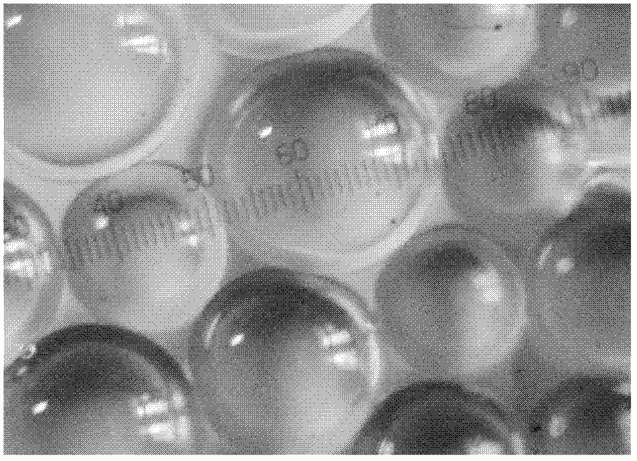
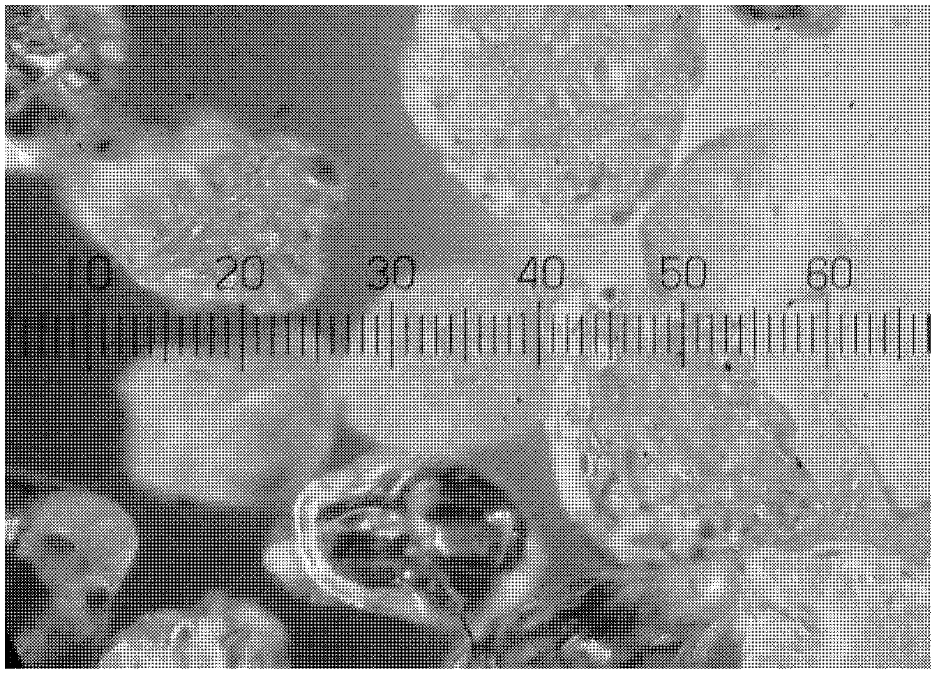
[0040] a. Morphological observation

[0041] The microspheres were soa...

Embodiment 2

[0050] Embodiment 2: Preparation of gelatin microspheres

[0051] (1) preparation concentration is the gelatin aqueous solution of 40% g / ml;

[0052] (2) Add the aqueous gelatin solution prepared in step (1) to liquid paraffin containing 2% g / ml Span 80 at 50°C, stir for 10 minutes, then drop the temperature to 2-5°C, stir for 30 minutes, and the stirring speed Be 350rpm, leave standstill, the microsphere that makes formation settles;

[0053] (3) pouring over to remove the oil phase, and repeatedly washing the oil phase remaining on the surface of the microspheres with acetone, and filtering out the microspheres;

[0054] (4) Add the microspheres that step (3) obtains to pH 8~9, concentration is the glutaraldehyde aqueous solution of 41.6% g / ml and stir for 5 minutes, after reacting for 40 hours at 4 DEG C, wash the microspheres with distilled water, Freeze-dried and stored.

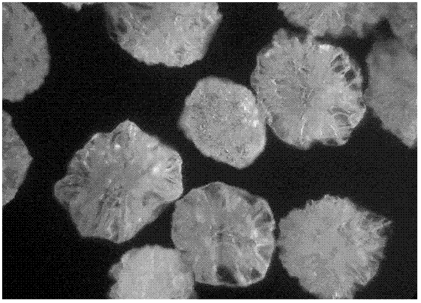
[0055] Observe the morphology of the microspheres in the same manner as in Example 1. The microsp...

Embodiment 3

[0059] Embodiment 3: Preparation of gelatin microspheres

[0060] (1) Take gelatin and prepare an aqueous gelatin solution with a concentration of 30% g / ml;

[0061] (2) Add the aqueous gelatin solution prepared in step (1) to liquid paraffin containing 0.8% g / ml Span 80 at 50°C, stir for 10 minutes, then drop the temperature to 2-5°C, stir for 30 minutes, and the stirring speed Be 300rpm, leave standstill, make the microsphere that forms settle;

[0062] (3) pouring over to remove the oil phase, and repeatedly washing the oil phase remaining on the surface of the microspheres with acetone, and filtering out the microspheres;

[0063] (4) Add the microspheres obtained in step (3) to pH 8-9 and formaldehyde solution with a concentration of 1.3% g / ml and stir for 5 minutes, react at 4°C for 13 hours, wash the microspheres with distilled water, and freeze-dry save.

[0064] Observe the morphology of the microspheres in the same manner as in Example 1. The microspheres in this ...


PUM

PropertyMeasurementUnit
particle diameteraaaaaaaaaa

Abstract

The invention provides a gelatin embolism microsphere and a preparation method and an application thereof. The preparation method of the gelatin embolism microsphere comprises the following steps of: (1) preparing a gelatin aqueous solution of which the concentration is 10-50 percent g / ml; (2) adding the gelatin aqueous solution prepared in the step (1) into an emulsifier-containing oil phase of which the temperature is 50-60 DEG C, stirring for 10 minutes, lowering the temperature to 2-5 DEG C, stirring for 30 minutes at the stirring speed of 100-900 revolutions per minute, and standing to precipitate the microsphere; (3) pouring the oil phase out, repeatedly washing the oil phase left on the surface of the microsphere with acetone, and filtering the microsphere out; and (4) adding the microsphere obtained in the step (3) into a crosslinking agent aqueous solution of which the pH is 8-9 and the concentration is 1-15 percent g / ml, reacting at the temperature of 4 DEG C for 0.5-25 hours, and washing the microsphere with distilled water. The gelatin embolism microsphere provided by the invention is crosslinked uniformly, is regularly spherical, and is convenient for screening grading and accurate marking of the particle diameter; and the degradation time and elasticity of the gelatin embolism microsphere are more suitable for clinical application.

Description

technical field [0001] The invention belongs to the field of interventional medicine, and relates to a gelatin embolization microsphere for embolization therapy, a preparation method and application thereof. Background technique [0002] Interventional therapy is one of the fastest growing medical disciplines in the 21st century. At present, it has kept pace with medical treatment and surgical treatment, and has become the third treatment system. Embolization therapy is an important part of interventional therapy, and it is a minimally invasive treatment using modern high-tech means. Embolization therapy is a local treatment that artificially blocks blood vessels by introducing embolic agents into the human body through precision instruments such as special catheters and guide wires under the guidance of medical imaging equipment. Embolization therapy has achieved good results in the treatment of malignant tumors, uterine fibroids, hemangiomas, vascular malformations, and ...

Claims


Application Information

Patent Timeline
Patent Type & Authority Applications(China)
IPC IPC(8): C08J3/24C08L89/00A61L31/04A61L31/14A61L31/16A61K9/16A61K9/19A61K47/42A61K45/00A61K49/00A61P35/00A61P7/04
Inventor 范田园吴雅楠崔代超张安阳张苑
Owner HYGEA MEDICAL TECH CO LTD
Why Eureka
Try Eureka