System for detecting breast cancer
A breast cancer and buffer technology, applied in the field of medicine, can solve the problems of quantification, inability to generate, increase the detection cost and detection difficulty, etc., and achieve the effect of simple operation and short detection time.
- Summary
- Abstract
- Description
- Claims
- Application Information
AI Technical Summary
Problems solved by technology
Method used
Image
Examples
Embodiment 1
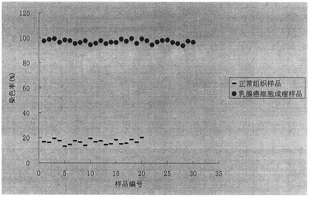
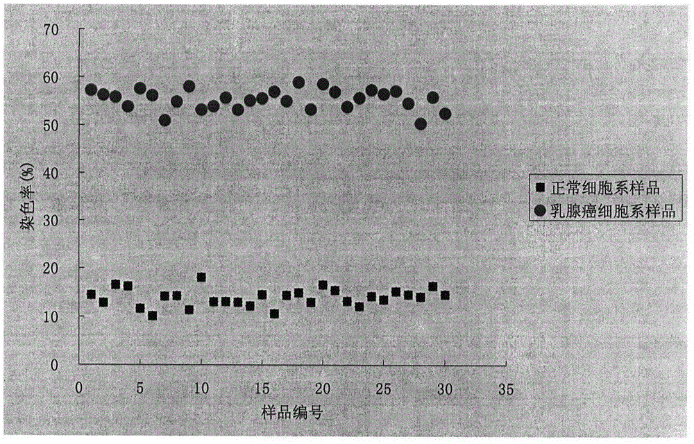
[0037] Feed 50 nude mice, 20 of which were used as blank controls, and 30 of them were inoculated with breast cancer cells (MCF-7) in the nude mice. After the tumors grew, tumor tissues were taken from the nude mice inoculated with MCF-7, and the blank control group was directly Take mammary gland and nearby muscle cells, make a single cell suspension, wash and suspend with sodium phosphate-sodium hydrogen phosphate buffer solution with a pH value of 6, and obtain 20 parts with a concentration of 10 4 ~10 9 cells / mL of normal cell suspension samples and 30 copies with a concentration of 10 4 ~10 9 cells / mL of tumor cell suspension samples.
[0038] Add the compound of the following formula to each sample so that the concentration of the compound in each sample is 0.5 μM
[0039]
[0040] Each sample was incubated in a 37°C incubator for 1 hour, followed by an additional 1 hour in a 4°C refrigerator.
[0041]After the incubation, the samples were centrifuged to remove th...
Embodiment 2
[0046] The same steps as in Example 1 were used to detect the tumor cells and normal cells of the above 50 nude mice, the difference being that the compounds used were:
[0047]
[0048] The concentration of the compound in the sample was 50 μM, and the incubation temperature and time were 15 minutes at 37° C. and 15 minutes at 4° C., and then the staining rate of each sample was obtained by using the fluorescence signal above 670 nm of the flow cytometer sample.
[0049] Test results: the staining rates of all breast cancer samples are above 90%, and the staining rates of all control samples are below 20%.
Embodiment 3
[0051] The same steps as in Example 1 were used to detect the tumor cells and normal cells of the above 50 nude mice, the difference being that the compounds used were:
[0052]
[0053] The compound concentration was 10 μM, the incubation temperature and time were 7.5 hours at 37°C, and 0.5 hours at 4°C, and the fluorescence signal above 670nm was detected by flow cytometry to obtain the staining rate of each sample.
[0054] Test results: the staining rates of all breast cancer samples are above 80%, and the staining rates of all control samples are below 25%.
PUM
 Abstract
Description
Claims
Application Information
 - R&D
- Intellectual Property
- Life Sciences
- Materials
- Tech Scout
- Unparalleled Data Quality
- Higher Quality Content
- 60% Fewer Hallucinations
Browse by: Latest US Patents, China's latest patents, Technical Efficacy Thesaurus, Application Domain, Technology Topic, Popular Technical Reports.
© 2025 PatSnap. All rights reserved.Legal|Privacy policy|Modern Slavery Act Transparency Statement|Sitemap|About US| Contact US: help@patsnap.com


