Preparation method and application of tissue slice for observing temporal-spatial distribution of early embryo development in vivo

A time-space distribution and tissue slicing technology, applied in the biological field, can solve the problems of inconvenient operation and observation, inability to reflect the positional relationship of embryos well, difficulty in popularization, etc., and achieve the effect of continuity

Inactive Publication Date: 2013-02-27
NORTHWEST A & F UNIV
View PDF5 Cites 15 Cited by
  • Summary
  • Abstract
  • Description
  • Claims
  • Application Information

AI Technical Summary

Problems solved by technology

Since mice are animals that develop in the uterus, the embryos are small in size and buried deep in the reproductive tract of the mother, which is not convenient for related operations and observations. Therefore, simple stereomicroscopic observation or combined with micromanipulation techniques cannot well reflect the development of embryos in the reproductive tract. The positional relationship of the differential interference phase contrast microscope is difficult to popularize and promote the use in general laboratories due to the high requirements for microscope operation technology.

Method used


Image

  • Preparation method and application of tissue slice for observing temporal-spatial distribution of early embryo development in vivo
  • Preparation method and application of tissue slice for observing temporal-spatial distribution of early embryo development in vivo
  • Preparation method and application of tissue slice for observing temporal-spatial distribution of early embryo development in vivo

Examples


preparation example Construction

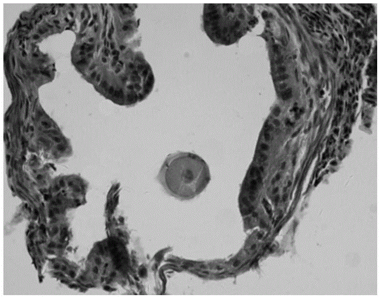
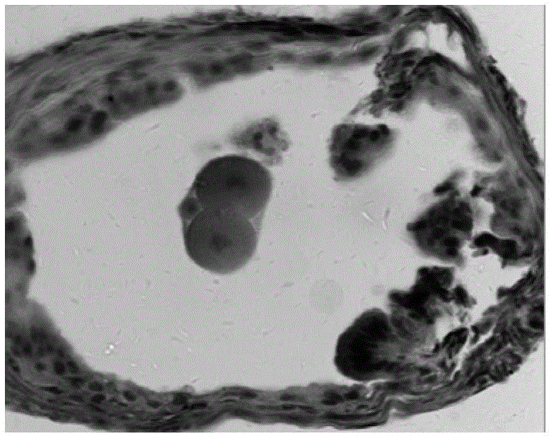
[0051] A method for preparing tissue slices for observing the temporal and spatial distribution of early mouse embryo development, comprising the following steps:

[0052] (1), mouse embryo model establishment and specimen collection

[0053] Select healthy Kunbai mice (200 female mice, weighing 25-30 g; 200 male mice, weighing 30-35 g). The room temperature was kept at 22°C, and the male mice were reared in a single cage under light-controlled conditions. Mice were used for experiments at least one week after they were purchased and acclimatized to the environment. Female mice were intraperitoneally injected with PMSG (10IU / mouse, Ningbo No. 2 Hormone Factory) at 5:00 p.m. 48 hours later, intraperitoneally injected with hCG (10IU / mouse, Ningbo No. 2 Hormone Factory), and mated with male rats (1:1) in the same night , Check the vaginal suppository the next morning. See Shuan as 0d, and the fallopian tubes covering fertilized eggs, 2-cell embryos, 4-cell embryos, and 8-cell ...

Embodiment 1

[0061] In vivo developmental time course and spatial position observation of early embryos of superovulated mice:

[0062] (1) Experimental animals and superovulation treatment: female Kunbai mice (6-8 weeks old, body weight 25-28 g) and male mice (8-12 weeks old, body weight 35-40 g) were selected. Under strict light control conditions (12h circadian rhythm), the male mice were reared in single cages. The female mice were intraperitoneally injected with 10IU PMSG at 15:00, and 10IU hCG was injected intraperitoneally at intervals of 48h for super-ovulation treatment. Immediately after the hCG injection, the female mice were placed in the same cage as the male mice overnight. In the morning of the next day, check whether the female rats have vulva plug formation, and those who are positive for the vulva plug inspection are presumed mated females, and they are picked out for use;

[0063] (2) Determination of the developmental time course of mouse embryos in vivo: based on the...

Embodiment 2

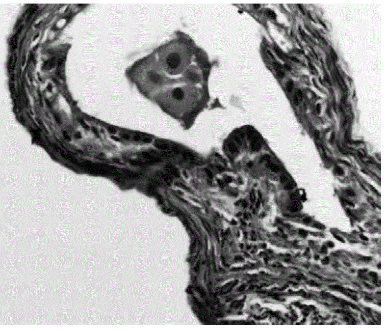
[0074] Crb3 expression in mouse embryos at various stages of development:


PUM

PropertyMeasurementUnit
melting pointaaaaaaaaaa

Abstract

The invention discloses a preparation method and an application of a tissue slice for observing temporal-spatial distribution of early embryo development in vivo. The preparation method comprises the following steps that 4% paraformaldehyde fixing, upward gradient ethanol dehydration, wax dipping, embedding, serial section, baking, dewaxing and downward gradient ethanol rehydration are performed in sequence on oviducts or uterine tissues which contain mice embryos in every period, and finally, after haematoxylin-eosin staining is performed on the tissues in the slice, neutral gum is used for sealing the slice, or after immunofluorescence histochemical staining is performed on the slice, a fluorescence resistant quenching sealing agent is used for sealing the slice. The tissue slice disclosed by the invention can be used for manufacturing a map of early mice embryo development and detecting the expression of Crb3 in the mice embryos in every period of development in vivo. The preparation method has the advantages that positions of all organs in the embryos can be relatively fixed, so that the position change of embryo cells in a genital tract and the continuity of embryo development can be conveniently observed, the structure is clear, and the tissue slice is convenient to store.

Description

technical field [0001] The invention belongs to the field of biotechnology, and in particular relates to a preparation method and application of tissue slices for observing the temporal and spatial distribution of early embryo development in vivo. Background technique [0002] In recent years, a series of embryonic engineering studies carried out on mice as mammalian model organisms, such as transgenic mice, gene knockout mice, cloned mice, and mouse embryonic stem cell engineering, have become commonly used research animals. It is based on the observation and operation of early mouse embryo development, so it is particularly important to be familiar with and master the embryo development time course and morphological structure changes. [0003] The commonly used method for studying the developmental time course of early embryos in mice is to use a stereo microscope to observe, and gross anatomy and micromanipulation techniques are also used to observe the developmental time...

Claims


Application Information

Patent Timeline
Patent Type & Authority Applications(China)
IPC IPC(8): G01N1/28G01N1/30
Inventor 卿素珠张为民徐永平杨媛媛袁苏雅徐金霞姬丽娜
Owner NORTHWEST A & F UNIV
Why Eureka
Try Eureka