
Testing method and kit for secondary circulation amplification of microRNA (Ribose Nucleic Acid)
A detection method, a quadratic technology, applied in biochemical equipment and methods, microbial measurement/inspection, etc., can solve the problems of complex PCR design, short miRNAs sequence, and low sensitivity
- Summary
- Abstract
- Description
- Claims
- Application Information
AI Technical Summary
Problems solved by technology
Method used
Image

Examples
Embodiment 1
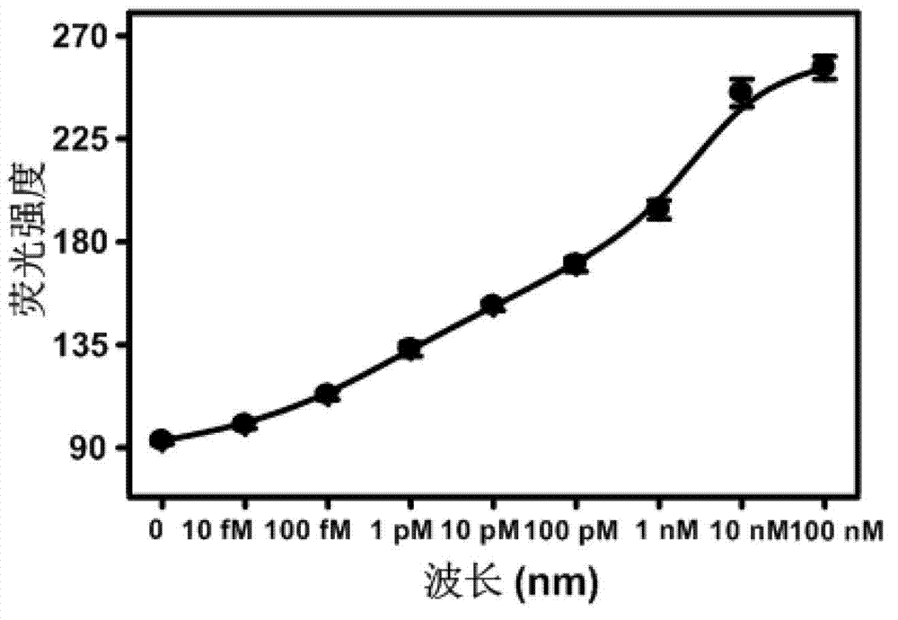
[0055] Example 1 uses miR-21 as a test object to further illustrate the present invention.
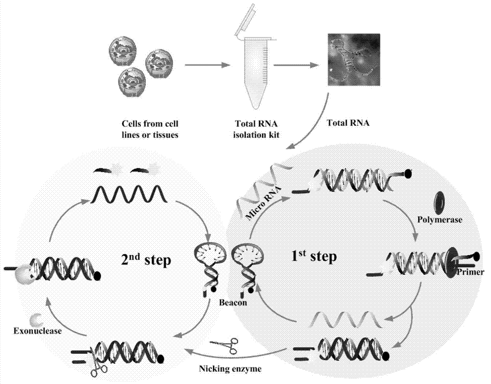
[0056] The enzymes and buffers used in this example include: NEB buffer 2, Bst large fragment DNA polymerase, lambda exonuclease, Nb.BbvCI nicking endonuclease, etc. were all purchased from NEB Company; molecular beacon probes, BSA, 8-base primers, miRNAs, DEPC water, dNTP, RNase inhibitor, etc. were purchased from Dalian Biotech.
[0057] The instrument used for fluorescence detection is a fluorescence spectrophotometer (Hitachi F-4500, Japan). Fluorescence spectrum measurement conditions: both the excitation and emission narrow peak widths are 5nm, the voltage PMT is 700v, the excitation wavelength is 490nm, and the emission wavelength scanning range is 500-600nm; a 250μL quartz cuvette is used for measurement.
[0058] The specific detection method is:
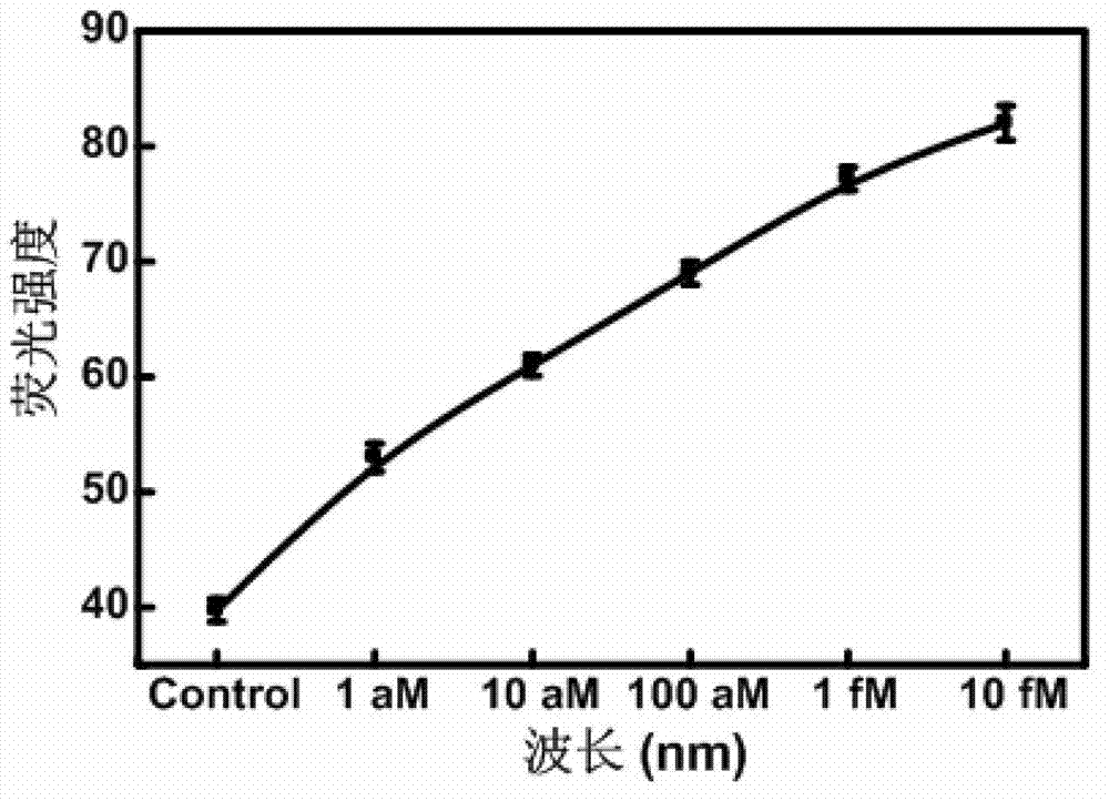
[0059] Step 1: Detection standard curve at 37°C: take 5-8 1.5ml centrifuge tubes, add molecular beacon probes with a final conce...
Embodiment 2
[0062] Example 2 uses the extract of breast cancer cells as the test substance to further illustrate the present invention.
[0063] Step 1: detection of breast cancer cells. Use mirVana first TM The kit extracts miRNA as the sample to be detected.
[0064] Step 2: Mix the sample to be tested with a final concentration of 500nM molecular beacon probe, 2μM 8-base primer, 400μM dNTPs, 16U Bst polymerase, 15U Nb.BbvCI nicking endonuclease, 12.5Ulambda exonuclease, Add BSA and RRI to a 1.5ml EP centrifuge tube. Detection was performed using a fluorometer. The breast cancer cell detection standard curve of cell number and fluorescence intensity can be obtained. (See Figure 4 ). The instrument setup parameters remain unchanged.
[0065] Step 3: We verified the extraction results of 1000 breast cancer cells. Using the above steps, we finally obtained a fluorescence value of 135. By comparing with the detection standard curve of breast cancer cells, it was found that 135 just...
PUM
 Abstract
Description
Claims
Application Information