
Preparation method of nano micrometer structure coexistence chitosan double-layer support

A micron structure, chitosan technology, applied in medical science, prosthesis, etc., can solve problems that cannot adapt to complex organs and tissue repair of the human body

Active Publication Date: 2014-09-10
QINGDAO UNIV
View PDF8 Cites 1 Cited by
  • Summary
  • Abstract
  • Description
  • Claims
  • Application Information

AI Technical Summary

Problems solved by technology

However, most of the chitosan biomaterials prepared at present have a single microstructure, which cannot adapt to the repair of complex organs and tissues of the human body. The environment in which bone cells grow

Method used


Image

  • Preparation method of nano micrometer structure coexistence chitosan double-layer support
  • Preparation method of nano micrometer structure coexistence chitosan double-layer support
  • Preparation method of nano micrometer structure coexistence chitosan double-layer support

Examples


Embodiment

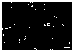
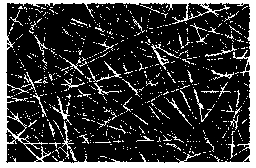
[0017] Embodiment: add 2.0g pure acetic acid in the distilled water of 98ml, mix well. Add 6g of chitosan powder to this acetic acid solution, dissolve with electromagnetic stirring, and form a uniform viscous solution after 12 hours. Pour the chitosan solution into a 24-well cell culture plate, vacuum out the air bubbles in the solution, and place it in a freezer at -18°C overnight to form a chitosan gel. Put the gel into a vacuum drier to freeze-dry for 3 days, take it out, and cut it into micron chitosan sheets with a thickness of about 2 mm. Paste micron chitosan flakes on aluminum foil with conductive double-sided adhesive tape. Weigh 0.7g chitosan powder, add 9.3g trifluoroacetic acid. Dissolve chitosan at 70°C under reflux conditions. Take 2ml of chitosan trifluoroacetic acid solution, add it to a 3ml needle tube, pass through 15kV high-voltage static electricity, the distance between the needle tube and the substrate micron chitosan sheet is 20cm, and the high-volta...


PUM

PropertyMeasurementUnit
porosityaaaaaaaaaa

Abstract

The invention discloses a method for preparing a chitosan double-layer scaffold with coexistence of nano and micro structures, which belongs to the field of biomedical materials. It is the first to use phase separation technology to prepare three-dimensional micro-chitosan scaffold as the base layer, and use high-voltage electrospinning technology to spin nano-chitosan fibers on the surface of micro-chitosan, so as to obtain chitosan bilayer with nano-micro structure coexistence. layer bracket. The prepared double-layer scaffold has good mechanical strength and high porosity, and is suitable for cell adhesion, growth and differentiation. Among them, the surface chitosan nanofiber is suitable for the growth of chondrocytes, and the base layer micron chitosan scaffold is suitable for the growth of osteoblasts, and can be applied to the repair of human cartilage and hard bone joints.

Description

technical field [0001] The technical field of the invention relates to biomedical materials, and relates to a preparation method of a chitosan bilayer bracket with coexistence of nano and micro structures. Background technique [0002] Tissue engineering is an interdisciplinary science that applies the principles of cell biology and engineering to research and develop biologically active substitutes for repairing or improving the structure and function of human diseased tissues or organs. Its basic principle and method are to induce differentiation and proliferation of autologous or allogeneic living cells, plant them on a biomaterial with excellent cytocompatibility and can be degraded and absorbed by the body to form a complex, and then combine the cell-biomaterial When biomaterials are implanted into the damaged parts of human tissues and organs, while the biomaterials are gradually degraded and absorbed by the body, the cells continue to proliferate and differentiate, fo...

Claims


Application Information

Patent Timeline
Patent Type & Authority Patents(China)
IPC IPC(8): A61L27/40A61L27/20
Inventor 楼涛汪学军
Owner QINGDAO UNIV
Learn More