Superpara magnetism polymeric microsphere magnetic resonance imaging (MRI) contrast agent and preparation method thereof

A magnetic resonance imaging and polymer technology is applied in the magnetic resonance imaging contrast agent and its preparation, the magnetic resonance imaging contrast agent constructed by superparamagnetic polymer microspheres and the preparation field thereof, so as to improve the stability, acid resistance and alkali resistance. , good application prospect, small effect of toxic and side effects

Active Publication Date: 2013-06-12
XIAMEN UNIV
View PDF9 Cites 8 Cited by
  • Summary
  • Abstract
  • Description
  • Claims
  • Application Information

AI Technical Summary

Problems solved by technology

[0004] In summary, the MRI contrast agents for superparamagnetic polymer microspheres, especially those with a core-shell structure and embe

Method used


Image

  • Superpara magnetism polymeric microsphere magnetic resonance imaging (MRI) contrast agent and preparation method thereof
  • Superpara magnetism polymeric microsphere magnetic resonance imaging (MRI) contrast agent and preparation method thereof
  • Superpara magnetism polymeric microsphere magnetic resonance imaging (MRI) contrast agent and preparation method thereof

Examples


Embodiment 1

[0028] Embodiment 1 Preparation of superparamagnetic polymer microsphere magnetic resonance imaging contrast agent

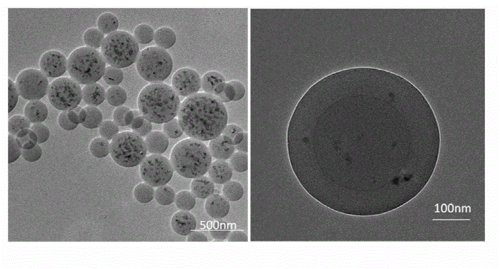
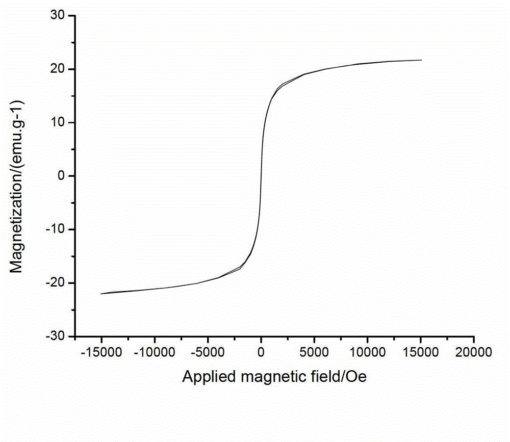
[0029] 5.41gFeCl 3 ·6H 2 O and 2.78g FeSO 4 ·7H 2 O was dissolved in 40mL of deionized water, and deoxygenated by nitrogen gas for 30min; 3.54g of NaOH was dissolved in 10mL of deionized water, added slowly, reacted at 60°C for 1h, added 2mL of oleic acid, and heated to 80°C for one hour to obtain 2-10nm particles. Magnetic Fe with superparamagnetism 3 o 4 Nanoparticles. The modified magnetic Fe 3 o 4 Nanoparticles were washed with ethanol for 2-3 times until neutral, and dried for later use. Take the prepared 0.4gFe 3 o 4 Nanoparticles, 2mL styrene, 0.04g isoalcohol diester maleate, and 40mL deionized water were fed at one time, and after stirring for 30 minutes, 0.02g ammonium persulfate was added and the temperature was raised to 65°C for 12~36h to obtain particles with a particle size of 200~300nm. Superparamagnetic polymer microspheres magnetic c...

Embodiment 2~9

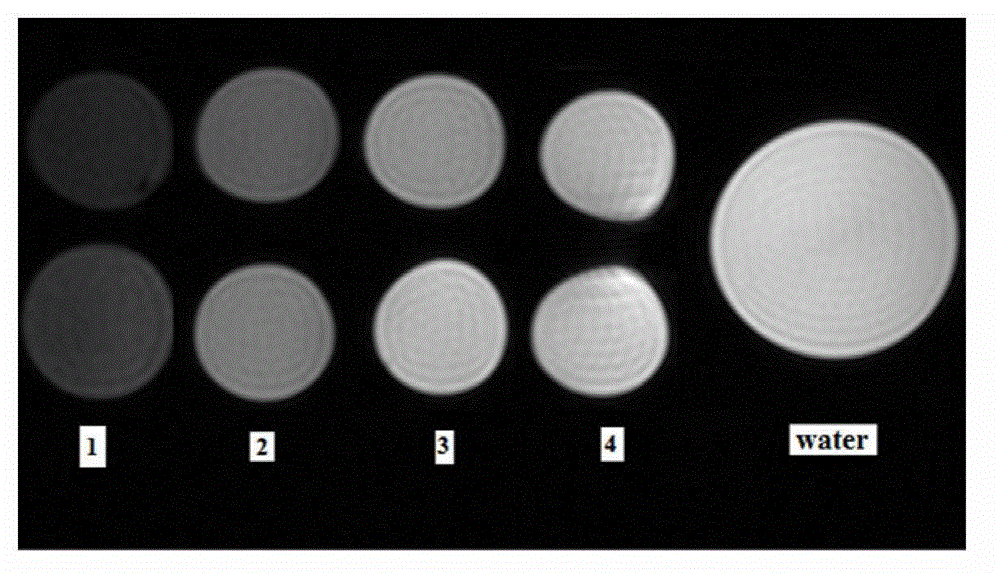
[0032] Using the same process as in Example 1, changing the feeding materials of styrene, isoalcohol diester maleate, and ammonium persulfate to prepare superparamagnetic polymer microsphere magnetic resonance contrast agents with different particle sizes, the results are shown in Table 1.

Embodiment 10

[0033]Example 10 Cytotoxicity Test of Superparamagnetic Polymer Microsphere Magnetic Resonance Imaging Contrast Agent

[0034] Implant Hela cells into 10mL cell culture dish bottles, the conditions are 37°C, saturated humidity, 5% CO 2 , the culture system is DMEM culture medium with superparamagnetic polymer microsphere magnetic resonance imaging contrast agent concentration of 100 μg / mL. The experimental results showed that the survival rate of the cells reached 89%, indicating that the toxicity and side effects were small.


PUM

PropertyMeasurementUnit
Particle sizeaaaaaaaaaa

Abstract

The invention discloses a superpara magnetism polymeric microsphere magnetic resonance imaging (MRI) contrast agent and a preparation method of the superpara magnetism polymeric microsphere MRI contrast agent, and relates to the MRI contrast agent used for diagnosis and treatment and the preparation method of the MRI contrast agent used for the diagnosis and the treatment. Polymeric microspheres are formed by polymerizable amphiphilic macromolecules, styrene monomers and Fe3O4 nano particles in a one-time batch feeding polymerization mode. Each microsphere comprises a core-shell structure, an inner core envelopes the superpara magnetism Fe3O4 particles, and a shell layer surface contains abundant functional groups. The microspheres are enabled to have targeting functions through coupling targeting molecules, and suitable for targeting MRI for certain diseases. The superpara magnetism polymeric microspheres are simple in synthetic process, low in preparation cost, controllable in microsphere particle diameter, stable in form, good in biocompatibility (BC) and MRI effect, and good in popularization and application values.

Description

technical field [0001] The invention belongs to the field of composite materials and molecular imaging, and relates to a magnetic resonance imaging contrast agent and a preparation method thereof, in particular to a magnetic resonance imaging contrast agent constructed of superparamagnetic polymer microspheres and a preparation method thereof. Background technique [0002] Magnetic resonance imaging (MRI) is one of the most powerful detection methods in clinical diagnosis today. It has the safety of no radiation damage, and can provide technical flexibility such as tomography in any direction, and can provide clear spatial resolution and physiological tissues and organs. Details. In MRI, because the relaxation times of some different tissues or tumor tissues overlap with each other, dynamic scanning and organ function measurement cannot be performed, and contrast agents are needed to improve imaging contrast. [0003] Magnetic resonance imaging contrast agents can generally...

Claims


Application Information

Patent Timeline
IPC IPC(8): A61K49/18A61K49/12
Inventor 戴李宗陈凌南曾碧榕康启龙高辉罗伟昂许一婷余世荣叶华立李聪陈国荣毛杰刘新瑜何凯斌
Owner XIAMEN UNIV
Why Eureka
Try Eureka