
Novel granulocyte preparation method and cancer-killing assay method of granulocytes

An anti-cancer activity, granulocyte technology, applied in biochemical equipment and methods, animal cells, vertebrate cells, etc., can solve the problems of killing, inability to find cancer, and unsatisfactory methods and effects of cancer treatment

Inactive Publication Date: 2013-07-24
JIANGSU ELEVATOR BIO TECH CO LTD
View PDF4 Cites 3 Cited by
  • Summary
  • Abstract
  • Description
  • Claims
  • Application Information

AI Technical Summary

Problems solved by technology

Although the research on cancer treatment at home and abroad has achieved phased results, the methods and effects of cancer treatment are still not satisfactory, and it is still impossible to find a treatment that can effectively kill, eliminate and reduce side effects on cancer

Method used


Image

  • Novel granulocyte preparation method and cancer-killing assay method of granulocytes
  • Novel granulocyte preparation method and cancer-killing assay method of granulocytes
  • Novel granulocyte preparation method and cancer-killing assay method of granulocytes

Examples


Embodiment 1

[0054] Inoculation of tumor cells

[0055] (1) will 1×10 4 Hela cells were inoculated in T25 culture flasks, with 3.5ml complete medium in each flask.

[0056] (2) At 37°C, 5% CO 2 , cultured in a saturated humidity incubator.

[0057] (3) Discard the culture medium in the T25 culture flask, add 5-10 ml of PBS to wash the cells, and discard the PBS by suction. Add 5 ml of 0.25% trypsin to digest the cells, observe the shrinkage and rounding of the cells under an inverted microscope, add 5 ml of medium to stop the trypsinization, transfer the cells into a 15 ml centrifuge tube with a pipette, take 0.1 ml for trypan blue staining and count the cells.

[0058] (4) Adjust the cell concentration to 1×10 6 cells / ml, the cells were added to the corresponding wells of the 96-well culture plate at 200 μl / well, 150 μl / well, 100 μl / well, 50 μl / well, and repeated wells. Add medium to the remaining experimental wells. Each well around the 96-well plate was filled with saline.

[005...

Embodiment 2

[0061] Peripheral blood collection and red blood cell precipitation

[0062] Aseptically collect 2ml of peripheral blood from the blood donor, anticoagulate with heparin, and fill in the relevant information of the blood donor on the blood collection tube. Use the precipitation solution (LIFT-001) formula developed by our company, mix according to the ratio of blood: precipitation solution = 5:1 (v / v), and let stand for 90 minutes. Shake 3 times during this time.

Embodiment 3

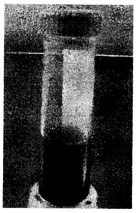
[0064] Preparation of granulocytes

[0065] Use the LIFT-002 separation liquid formula developed by our company to prepare 50ml of 3 kinds of density granulocyte separation liquid

[0066] Granulocyte separation fluid name Granulocyte isolation master solution A Granulocyte isolation master solution B Granulocyte isolation master solution C Granulocyte Separation Medium 1 30 5 15 Granulocyte Separation Medium 2 26 6 18 Granulocyte separation medium 3 21 7 22

[0067] (1) Take three 50ml sterile centrifuge tubes, marked as 1, 2, and 3 respectively corresponding to granulocyte separation solution 1-3.

[0068] (2) Put the centrifuge tubes on the balance and weigh them respectively, and record the weight of each tube.

[0069] (3) Add granulocyte separation mother solution C and B to No. 1-3 centrifuge tubes according to the amount marked in the above table, and shake gently. Then add granulocyte isolation mother solution A a...


PUM


Abstract

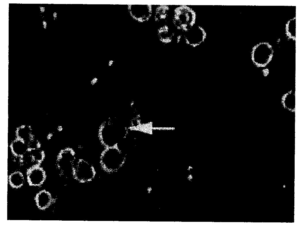
The invention relates to a granulocyte separating and purifying method, and especially relates to a novel granulocyte preparation method without damaging the activity of granulocytes, and a cancer-killing assay (CKA) method of the granulocytes. The CKA method comprises the following steps: 1, acquiring 2ml of the peripheral blood of a blood donor, and carrying out heparin anticoagulation; 2, mixing the blood with a precipitation liquid prepared through using a precipitation liquid (LIFT-001) formula developed by the applicant according to a volume ratio of the blood to the precipitation liquid of 5:1; 3, transferring the supernatant of a mixture obtained in step 2 to a centrifuge tube added with LIFT-002, centrifuging, and taking a buffy coat; 4, adding a culture medium, and counting for obtaining effector cells; incubating the effector cells and target cells; and 5, carrying out CKA detection. CKA experiment results show that the granulocytes of some healthy adults can effectively kill Hela tumor cells. Cell real-time monitoring system results show that cancer cells are broken and killed by the granulocytes. Cancer killing activity assay allows efficient donors having high activities to be screened, and can be used for the cancer prevention and treatment, and the management of dangerous factors.

Description

(1) The field of technology [0001] The present invention involves a method of separating and purified granulocytes, especially a new type of preparation method and anti-cancer activity detection method (CKA). (2) Technical background [0002] At present, cancer has become the first killer of human health, and has taken the precious life of millions of people.According to the latest information of the World Health Organization, about 6.6 billion people around the world, about 10 million new tumor patients each year, about 5 million people died of cancer, and a cancer patient died almost every 6 seconds. [0003] Its terribleness lies not only in the severe damage of the disease to the patient's body tissue and the high mortality rate of cancer patients, but also in traditional therapy, such as radiotherapy and chemotherapy, unbearable side effects.Although the research on the treatment of cancer at home and abroad has achieved staged results, the method and effect of treating canc...

Claims


Application Information

Patent Timeline
IPC IPC(8): C12Q1/18C12N5/0787
Inventor 王申俊崔征
Owner JIANGSU ELEVATOR BIO TECH CO LTD
Learn More