Method for distinguishing cancer cells through surface enhanced Raman spectroscopy

A surface-enhanced Raman, cancer cell technology, applied in the field of biomedicine, can solve the problems of scintillation, SERS spectral type peak position, peak intensity and peak number fluctuations, etc.

Inactive Publication Date: 2014-01-01
FUJIAN NORMAL UNIV
View PDF0 Cites 11 Cited by
  • Summary
  • Abstract
  • Description
  • Claims
  • Application Information

AI Technical Summary

Problems solved by technology

[0004] However, like other cell SERS detection methods, the cell SERS spectrum obtained by the electroporation cell SERS detection method also has a "flickering" phenomenon, that is, withi

Method used


Image

  • Method for distinguishing cancer cells through surface enhanced Raman spectroscopy
  • Method for distinguishing cancer cells through surface enhanced Raman spectroscopy
  • Method for distinguishing cancer cells through surface enhanced Raman spectroscopy

Examples


Embodiment 1

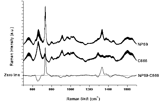
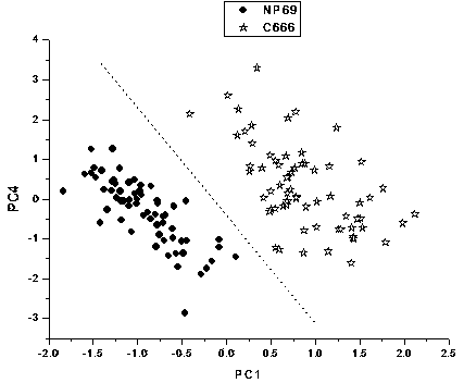
[0043] Distinguishing between normal nasopharyngeal NP69 cells and nasopharyngeal carcinoma C666 cells by surface-enhanced Raman spectroscopy

[0044] Take 100 μl and 400 μl of silver sol and normal nasopharyngeal NP69 cells or nasopharyngeal carcinoma C666 cells, mix them at room temperature, and transfer them into an electric shock cup. The electrode gap of the electric shock cup is 1 mm, and the number of cells is about 4000. Place the electric shock cup in ice bath for 5 minutes, ultrasonic pretreatment for 30 seconds, then place it in the electroporator, select manual control, apply a voltage of 350V, and last for 100ms. After electroporation, use 400μl RPMI 1640 cell culture medium to wash out the cells immediately After placing the sample pool in an ice bath for 5 minutes, transfer it to a preheated incubator, incubate at 37°C for 10 minutes, and then use a confocal Raman spectrometer for single-cell surface-enhanced Raman spectroscopy detection. The wavelength of the l...

Embodiment 2

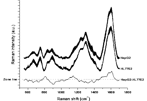
[0046] Differentiation of normal liver HL7702 cells and liver cancer HepG2 cells by surface-enhanced Raman spectroscopy

[0047] Take gold sol, 250 μl and 650 μl of normal liver HL7702 cells and liver cancer HepG2 cells, mix them at room temperature, and transfer them into an electric shock cup. The electrode gap of the electric shock cup is 4 mm, and the number of cells is about 6000. Place the electric shock cup in ice bath for 8 minutes, ultrasonic pretreatment for 60 seconds, then place in the electroporation instrument, select manual control, apply a voltage of 880V, and last for 50 ms. After electroporation, wash the cells with 800 μl RPMI 1640 cell culture medium immediately After taking out the sample pool, put it in an ice bath for 8 minutes, transfer it to a preheated incubator, and incubate at 37°C for 15 minutes, and then use a confocal Raman spectrometer to perform single-cell surface-enhanced Raman spectroscopy detection. The wavelength of the laser used to detec...


PUM


Abstract

The invention provides a method for distinguishing cancer cells through surface enhanced Raman spectroscopy. According to the method, after SERS active nanometer materials and various cells incubated under an ice-bath condition are rapidly preprocessed through ultrasonic, the SERS active nanometer materials are rapidly guided into the various cells through an electroporation method, the surface enhanced Raman spectroscopy of cancer cells is obtained by means of the detection of a Raman spectrometer, a surface enhanced Raman spectroscopy database of the various cells is built, clustering analysis is carried out through multivariate statistics analysis, a distinguishing splattering distribution graph corresponding to the surface enhanced Raman spectroscopy of normal cells and the cancer cells is obtained, and distinguishing of the cancer cells is achieved according to the distinguishing splattering distribution graph. The method for distinguishing the cancer cells through the surface enhanced Raman spectroscopy has the advantages of being rapid and easy to operate, good in universality, low in cost, free of flickering cell SERS spectrums and the like, can achieve large-scale cancer cell detection, and is suitable for being widely used in the technical field of medicine screening, disease diagnosis and the like.

Description

technical field [0001] The invention relates to a cancer cell surface-enhanced Raman spectrum discrimination method. Specifically, cancer cells and nanomaterials with SERS activity are pretreated with a pulsed electric field to detect surface-enhanced Raman spectrum signals of cancer cells, and finally use multivariate Analytical techniques Methods for performing statistical analysis and discrimination. Belongs to the field of biomedicine. Background technique [0002] Raman spectroscopy (Raman spectroscopy) is a molecular spectroscopy technology developed based on the Raman scattering effect. Using Raman spectroscopy technology can detect biological molecules such as proteins, nucleic acids, lipids, and sugars in tissue cells at the molecular level. fine structure and information. In the process of tumor growth and development, the material structure, conformation and quantity of the above molecules will change significantly. By comparing the Raman spectra of cancerous an...

Claims


Application Information

Patent Timeline
IPC IPC(8): G01N21/65
Inventor 林居强陈荣冯尚源陈冠楠黄祖芳俞允陆鹏
Owner FUJIAN NORMAL UNIV
Why Eureka
Try Eureka