Preparation method of biological wound protecting film

A biological and fetal membrane technology, applied in medical science, bandages, prostheses, etc., can solve the problems of expensive treatment, complex artificial skin, patient pain, etc., to achieve simple and easy-to-use equipment, uncomplicated preparation process, and no allergic reactions. Effect

Inactive Publication Date: 2014-03-12
曲凯明
View PDF6 Cites 6 Cited by
  • Summary
  • Abstract
  • Description
  • Claims
  • Application Information

AI Technical Summary

Problems solved by technology

Not only the cost of treatment is expensive, but more importantly, it brings great pain to patients
In addition, for patients with extensive b...

Method used


Image

  • Preparation method of biological wound protecting film
  • Preparation method of biological wound protecting film
  • Preparation method of biological wound protecting film

Examples


Embodiment 1

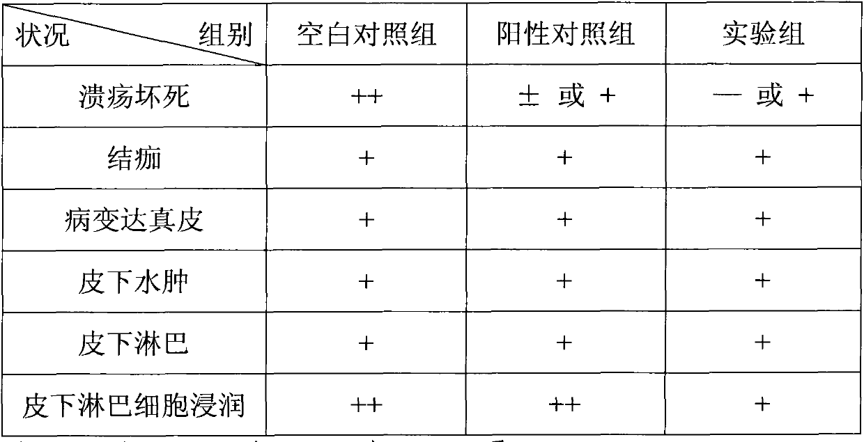
[0042] Embodiment 1: to the therapeutic effect of scald and pathological examination result

[0043] 36 mice weighing 20-24 g were randomly divided into 3 groups, 12 mice per group; the back hair was depilated with 10% sodium sulfide, and II degree scald surface with an area of ​​1.4×1.4 cm was caused with 80°C water. The first group of mice was treated with biological wound protection film as the experimental group; the second group of mice was treated with Jingwanhong scald medicine, which was used as the positive control group, and the third group of mice were used with everything, as the blank control group.

[0044]The results showed that: 1. The experimental group and the positive control group began to grow scabs on the second day. After 4-7 days, the scabs on the skin of all mice came off, and the new granulation was ruddy, and hairs gradually grew. 2. The positive control group (i.e. the first group) is slower than the experimental group (i.e. the second group); 3. Th...

Embodiment 2

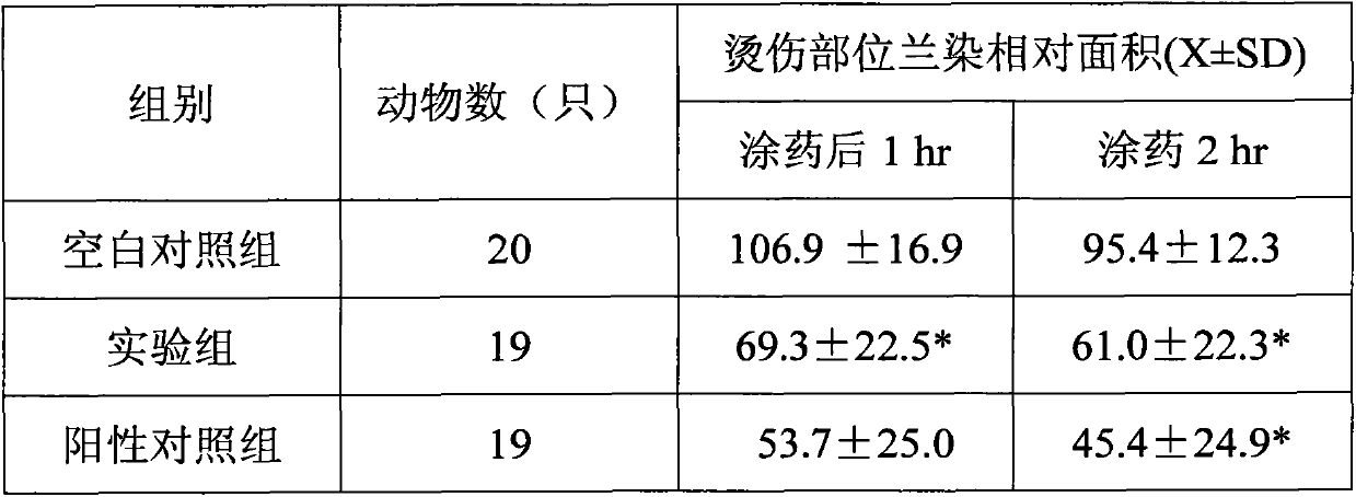
[0049] Example 2: Effects on Vascular Permeability of Scalded Rats

[0050] Take 20 white rats weighing 180-200g, anesthetize them by intraperitoneal injection of 1g / kg Uradum, fix the back, wash with 10% sodium sulfide abdominal hair removal normal saline and wait until dry, inject 1% Evans blue 0.2mL / rat sublingually Immediately use 80°C water to mark three symmetrical scald surfaces of 1.4×1.4cm on the abdomen, one is the blank control group (no medicine applied), one is the experimental group (that is, the place where the biological protective film is used), and one is the positive control In the group (i.e. using Jingwanhong scald medicine), the degree of blue staining on the scalded surface was observed after 1 hour and 2 hours after the scald medicine was applied or the wound film was applied, and the blue stained area was measured and calculated. The results are shown in the table below:

[0051]

[0052] *P<0.01 compared with the control group

[0053] It can be s...

Embodiment 3

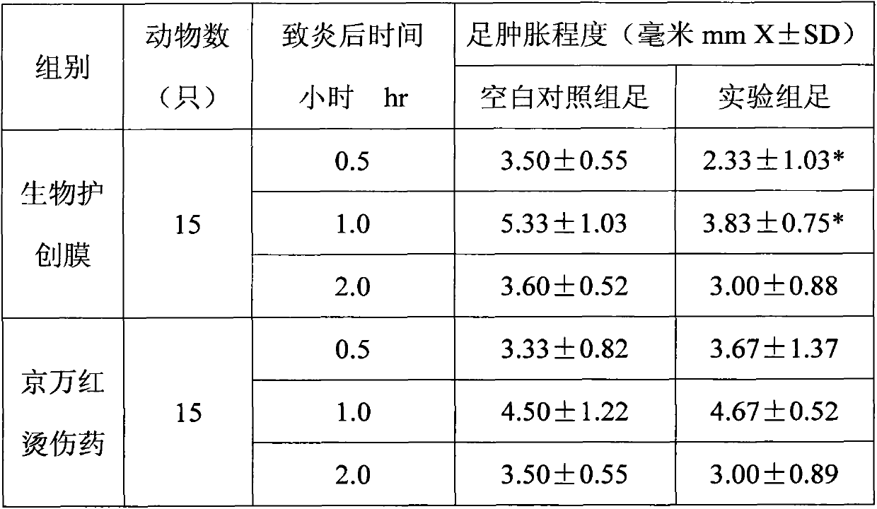
[0054] Example 3: Anti-inflammatory effect

[0055] Thirty white rats weighing 200-220 g were randomly divided into 2 groups, 15 rats per group, and the circumference of the two hind ankle joints and soles were measured. 20% fresh egg 0.1mL / foot was subcutaneously injected from the palmar aponeurosis of the foot to the ankle joint, and the right hind foot was used as the experimental group, in which the right hind foot of one group of rats was immediately pasted with biological wound film, and the right hind foot of the other group of rats was Immediately apply Jingwanhong scald medicine, and repeat the film or medicine application every 0.5hr. The left hind feet of all the rats in the above two groups are used as blank control group. At 0.5hr, 1hr, and 2hr after egg white injection, the two hindfoot joints and the meridians of the soles were re-measured, and the degree of inflammation and swelling was calculated. The results are shown in the table below:

[0056]

[0057]...


PUM


Abstract

The invention relates to a preparation method of a biological wound protecting film. The preparation method comprises the steps of (1) selecting a placenta, and taking down the caul; (2) washing to remove the chorion to obtain the amnion; (3) washing the amnion by normal saline, and soaking; (4) scrapping the amnion to remove the residual chorion and the appendage of the chorion; (5) washing; (6) putting the amnion into soak solution, treating and then fixing the amnion on a special diaphragm for standing; (7) washing the amnion by normal saline, and then washing the amnion by protecting liquid; (8) putting the amnion into clean protecting liquid, and irradiating by an ultraviolet lamp; (9) putting the amnion into a packaging bag, injecting the protecting liquid into the packaging bag and sealing to obtain the biological wound protecting film. The wound protecting film has the characteristics of being good in toughness, elasticity and air permeability, and the like; furthermore, the wound is not exclusive with the biological wound protecting film, and the biological wound protecting film is strong in application property; according to the wound protecting film and the preparation method of the wound protecting film, the cost is low, so that the financial burden and the pain of a patient are relieved, and dressing change and excision of eschar do not need to be carried out on a patient who has burn within three degrees; therefore, the biological wound protecting film is a most advanced biotechnology in China at present.

Description

technical field [0001] The invention relates to a medical material in the field of medicine, in particular to a preparation method of a biological wound protection film. Background technique [0002] Skin injuries caused by flames, hot solids, chemicals, intense radiant heat, and electricity are called burns. The degree of burn is different by the height of temperature, the length of action time. When the burned skin reaches more than 1 / 3rd of the whole body surface area, it can be life-threatening. [0003] Clinically, the severity of burn depends on the scope and depth of injured tissue, and the depth of burn can be divided into I degree, II degree and III degree. First-degree burns are the lightest, with redness, pain, obvious tenderness, oozing or edema on the burned skin, and partial whitening when the injured part is lightly pressed, but no blisters; second-degree burns are deeper, with blisters on the skin and the bottom of the blisters It is red or white, filled w...

Claims


Application Information

Patent Timeline
IPC IPC(8): A61L15/40A61L15/44A61L27/36A61L27/54A61L27/60
Inventor 汪洋李国义
Owner 曲凯明
Why Eureka
Try Eureka