

Intervertebral disc tissue scaffold including growth factor and preparation method thereof
A tissue scaffold and growth factor technology, applied in medical science, prosthesis, etc., can solve the problems of poor mechanical properties of hydrogel, easy cell death, and limited use, and achieve simple and easy preparation methods, good uniformity, and good The effect of mechanical properties
- Summary
- Abstract
- Description
- Claims
- Application Information
AI Technical Summary
Problems solved by technology
Method used
Image

Examples
Embodiment 1
[0027] Embodiment 1 adopts the method of the present invention to prepare the same intervertebral disc tissue support
[0028] Step 1. Dissolve polylactic acid-glycolic acid copolymer (PLGA) in hexafluoroisopropanol (HFIP) to make a 15% PLGA solution, which is the outer layer solution of coaxial electrospinning;
[0029] Step 2. Dilute TGF-β1 (Prospec Company) with 1% bovine serum albumin (BSA) to a concentration of 1 ng / μL;
[0030] Step 3. Dissolve polyvinyl acetate (PVA) in ultrapure water to form a 15% PVA solution.
[0031] Step 4. Add 6 μL of 1 ng / μL TGF-β1 to 2 mL of 15% PVA solution, which is the inner layer solution of coaxial electrospinning;
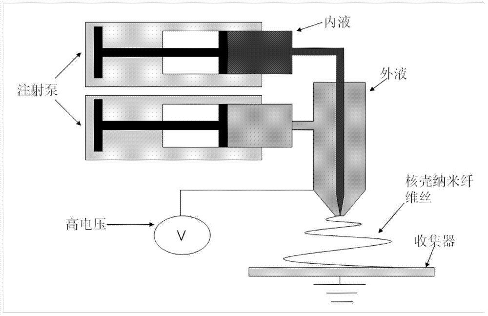
[0032] Step 5, such as figure 1 Assemble the instrument, the syringe is (LSP02-1B, Baoding Lange Company, China), the high-voltage power supply is an intermediate frequency DC high voltage device (RXZGF, Shanghai Rixing Company, China), the distance between the needle and the receiving device is 15cm, the voltage is 16-18Kv,...
Embodiment 2
[0034] Embodiment 2 detects the performance of the intervertebral disc tissue support of the present invention
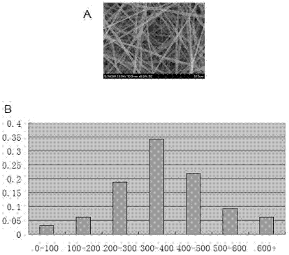
[0035] 1. Observing the constructed intervertebral disc tissue scaffold with a scanning electron microscope, and counting the distribution of spinning diameters. see results figure 2 . Scanning electron microscope results show that the spinning diameter is relatively uniform, and most of them are distributed in the range of 200-500nm. The double-layer structure of the intervertebral disc tissue scaffold can be clearly seen by the transmission electron microscope.
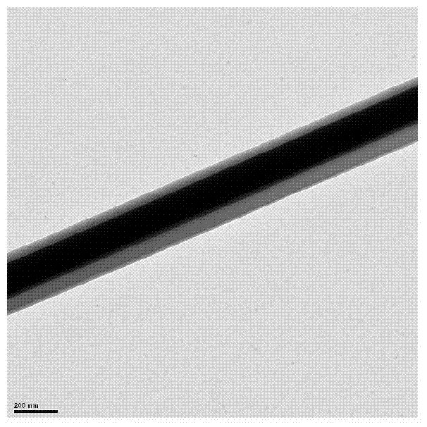
[0036] 2. Use a transmission electron microscope to observe the spinning shell-nucleus double-layer structure of the intervertebral disc tissue scaffold. The results are shown in image 3 . A clear boundary can be seen between the inner core layer and the outer shell layer, indicating that the spinning of inner and outer double shell-core structures was successfully obtained.
[0037] 3. In order to...
PUM
 Abstract
Description
Claims
Application Information