A fluorescent negative staining method for detecting living cells

A negative staining method and live cell technology, which is applied in the direction of fluorescence/phosphorescence, preparation of test samples, material excitation analysis, etc., can solve the problems of cell loss of viability, inapplicability of sampling and detection of precious cell samples, and impossibility of dynamic observation, etc. Achieve the effect of maintaining vitality and easy and convenient dyeing

Active Publication Date: 2017-10-20
江苏禄亿生生物科技有限公司
View PDF1 Cites 0 Cited by
  • Summary
  • Abstract
  • Description
  • Claims
  • Application Information

AI Technical Summary

Problems solved by technology

However, after Prussian blue staining, the cells lose their vitality, which is not suitable fo

Method used


Image

  • A fluorescent negative staining method for detecting living cells
  • A fluorescent negative staining method for detecting living cells
  • A fluorescent negative staining method for detecting living cells

Examples


Embodiment 1

[0031] Example 1: SPIO labeling mouse macrophage RAW264.7 cells

[0032] Take well-grown RAW264.7 cells, add SPIO (final concentration: 25-50 μg / ml) into the cell culture system, and place at 37°C, 5% CO 2 After incubation in an incubator for 6-24 hours, the labeling rate of SPIO on cells is close to 100%, and has no adverse effects on cell viability, growth, differentiation and other biological activities, and has no obvious cytotoxicity.

Embodiment 2

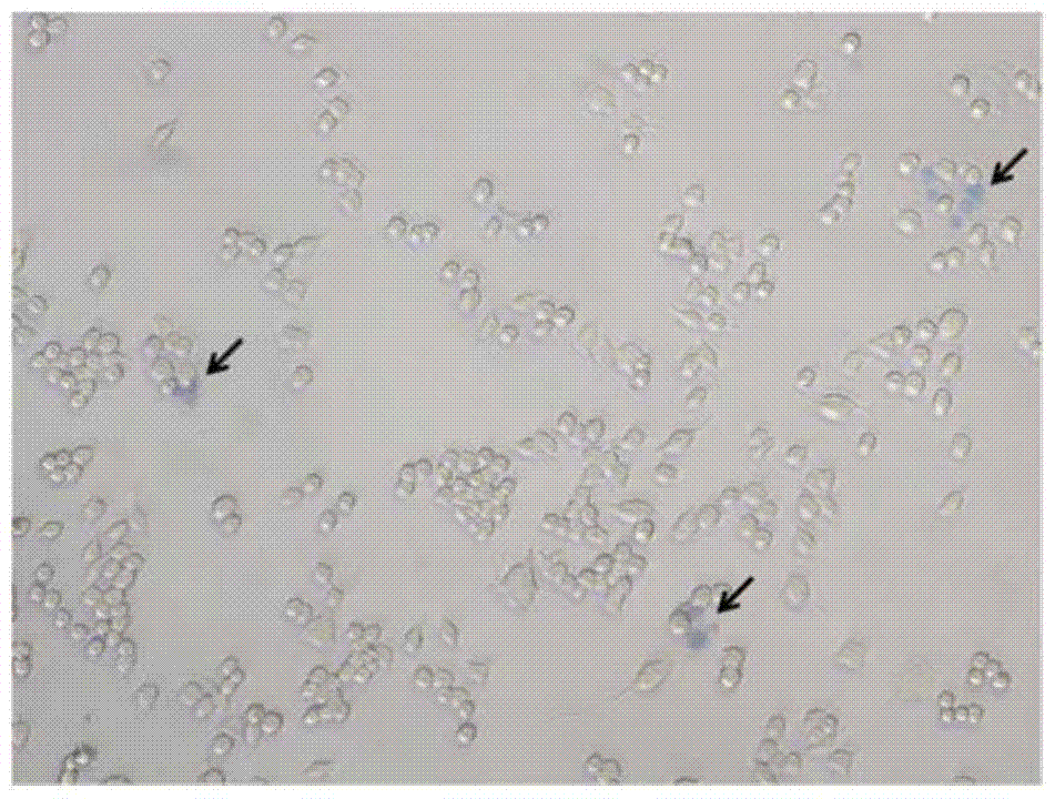
[0033] Embodiment 2: the Prussian blue staining method of cell slide

[0034] Place the sterile coverslip in a six-well culture dish, add SPIO-labeled RAW264.7 cells to prepare cell slides, fix with 4% paraformaldehyde for 10 minutes; wash with distilled water twice, each time for 2 minutes, and then dry in the air Dry; put the cell slide into Perls solution (preparation: 25ml of 20% potassium ferrocyanide aqueous solution, 25ml of 2% hydrochloric acid aqueous solution. The two solutions are prepared and stored separately, mixed in equal amounts before use, and used after filtration) for 30 minutes; distilled water Fully rinse; 0.2% Nuclear Fast Red (preparation: dissolve 10g of aluminum sulfate in 100ml of distilled water, add 0.2g of Nuclear Fast Red after dissolving, mix well, and put it in a 37°C water bath for 1 hour, shake it from time to time to fully dissolve it Filtered for use) counterstained for 20-30 minutes, rinsed with distilled water, and dried. Judgment of sta...

Embodiment 3

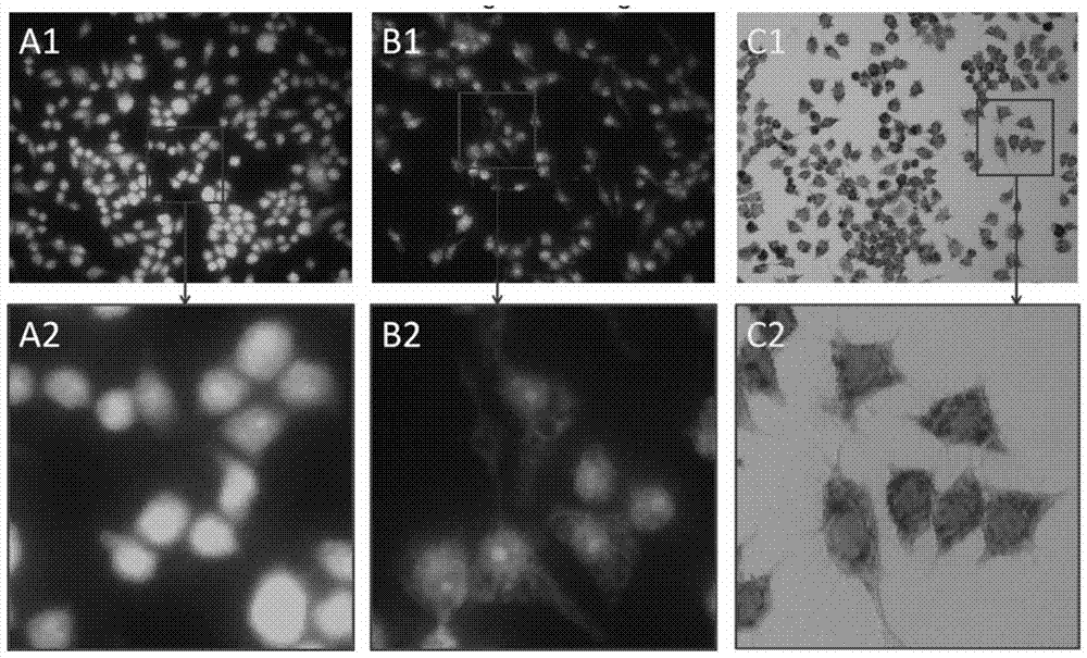
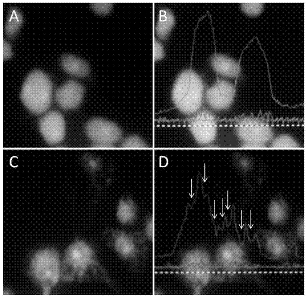
[0036] Example 3: CFSE fluorescent negative staining detects SPIO uptake by living cells

[0037]Prepare the SPIO-labeled RAW264.7 cell suspension, and adjust the cell concentration to about 107 cells / ml with an appropriate medium. Take 1ml of cell suspension into a test tube, add an appropriate amount of CFSE working solution, mix gently (the final concentration of CFSE is set to 0.1-2μM, and the optimal staining concentration is determined by the results of the preliminary experiment), and culture at 37°C Incubate in the box for 15-30 minutes. Remove the supernatant after centrifugation, add 2ml of PBS solution, remove the supernatant after centrifugation, and repeat this operation once. Add appropriate medium to make cell suspension. Drop 10 μl of cell suspension on a glass slide and observe with a fluorescent microscope. Results criteria: Negative (-), no fluorescent negative staining area; positive (+), including 1-2 fluorescent negative staining areas; positive (++), ...


PUM


Abstract

The invention discloses a method for detecting living cells by fluorescent negative staining. Fluorescent staining reagents stain whole cells, and when cells phagocytize opaque nano-particles, the nano-particles are enriched in the phagocytic body. Cut off the detection light so that it appears as a fluorescent negative staining area on the fluorescence image. According to the size of the negative staining area, the phagocytosis rate of the cells to the nanoparticles can be easily judged without affecting the activity of the cells. The CFSE fluorescence negative staining method of the cell labeling detection method of the present invention can be used for the detection of labeling rate and labeling amount after nano-iron-labeled cells, and for evaluating the pharmacodynamics and pharmacokinetics analysis of nano-iron-type new drugs, and the method Cells do not need to be fixed, CFSE staining is simple and convenient, and maintains the viability of cells.

Description

technical field [0001] The invention relates to the field of biotechnology, in particular to a method for detecting cell markers, in particular to a method for dynamically detecting phagocytosis of superparamagnetic iron oxide nanoparticles by a single living cell. Background technique [0002] With the rapid development of nanomaterial research, cell magnetic labeling has become a research hotspot in the field of imaging in recent years. Superparamagnetic iron oxide (SPIO) nanoparticles (superparamagnetic iron oxide, SPIO) is a new type of magnetic resonance contrast agent, added to the cell culture system in vitro, can be taken up by cells, and can be used for in vivo tracing after transfusion. Observing the migration and colonization of transplanted cells in various damage repair studies has been widely used in the research of mesenchymal stem cells, umbilical cord blood stem cells, and neural stem cells. SPIO is safe to use at a certain concentration, does not affect th...

Claims


Application Information

Patent Timeline
IPC IPC(8): G01N21/64G01N1/30
Inventor 何向锋强福林沈康蔡晶张一心王建红刘继斌
Owner 江苏禄亿生生物科技有限公司
Why Eureka
Try Eureka