A 3D printed porous metal with bionic three-dimensional micro-stent and its preparation method

A three-dimensional, three-dimensional image technology, applied in the field of biomedical materials, can solve the problems of inability to achieve growth, limited bone extraction, and inability to achieve scaffold bone volume.

Active Publication Date: 2017-04-12
PEKING UNION MEDICAL COLLEGE HOSPITAL CHINESE ACAD OF MEDICAL SCI
View PDF3 Cites 0 Cited by
  • Summary
  • Abstract
  • Description
  • Claims
  • Application Information

AI Technical Summary

Problems solved by technology

[0002] Bone defect treatment has always been a clinical problem. Traditional bone graft materials include autologous bone and allogeneic bone, but each has its own defects: autologous bone is usually taken from the patient's own ilium and fibula, which will not only cause trauma, infection, Moreover, the amount of bone taken is also limited; allogeneic bone has the risk of immune rejection and disease transmission
[0005] However, the 3D printed porous titanium alloy scaffold itself has no biological activity, and there are no sites that cells can recognize, and the various 3D printed porous titanium alloy materials currently on the market usually have large pore diameters, about 300-1500um in diameter, and an average of 20 μm for cells. The -30um diameter is obviously too open, and the cells can only grow in the two-dimensional space of the hole wall, and cannot achieve three-dimensional growth in the entire hole, and thus cannot achieve sufficient bone growth into the scaffold.
Previous studies have also focused on the two-dimensional activation modification of the inner surface of porous titanium pores, such as giving the surface acid-base treatment, surface plasma spray coating, surface loading growth factors, etc. Climbing, growth and matrix secretion and filling

Method used


Image

  • A 3D printed porous metal with bionic three-dimensional micro-stent and its preparation method
  • A 3D printed porous metal with bionic three-dimensional micro-stent and its preparation method
  • A 3D printed porous metal with bionic three-dimensional micro-stent and its preparation method

Examples


Embodiment 1

[0055] Example 1 Preparation of titanium alloy-gelatin / HA three-dimensional micro-stent

[0056] Preparation of titanium alloy-gelatin / HA three-dimensional microscaffold:

[0057] 1) Import the CT image into Mimics or CAS 3D image software to obtain a 3D image of the target bone tissue, fill and expand the image with a regular dodecahedral structural unit with a pore column of 300um and a pore diameter of 1500um, and obtain a personalized porous connected 3D image digital model;

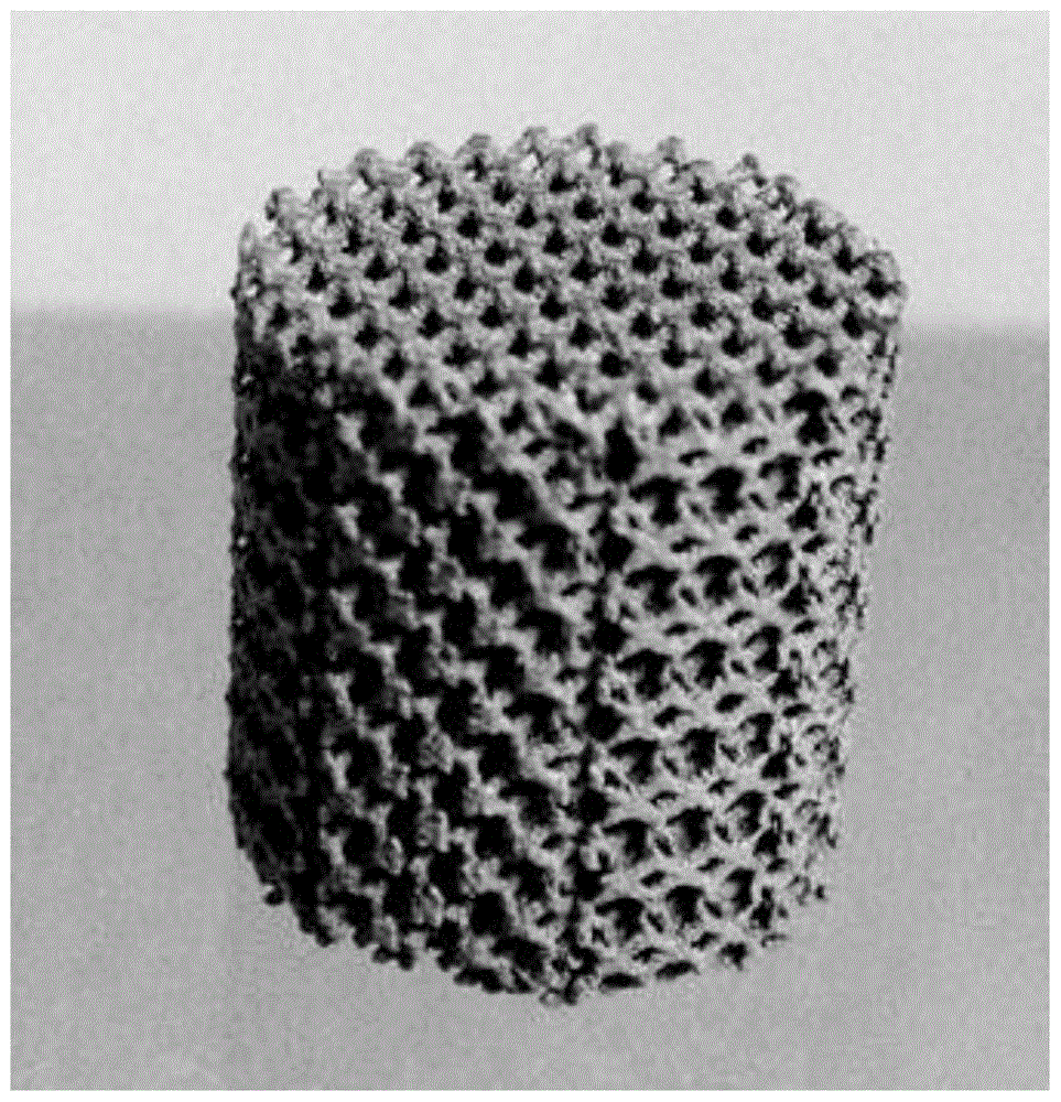
[0058] 2) Using EOS M280 metal material 3D printer, using titanium alloy as raw material, printing porous titanium scaffold according to the design model (see figure 1 );

[0059] 3) Add gelatin particles into deionized water at a ratio of 5%, and dissolve them completely in a magnetic stirrer at 37°C and 300r / min;

[0060] 4) Add nano-hydroxyapatite powder nHA to the gelatin solution at a ratio of 1:2 between nHA and gelatin, and use an ultrasonic cell pulverizer to disperse the mixture to a homo...

Embodiment 2

[0065] The characterization of embodiment 2 three-dimensional micro-stent

[0066] 1. Determination of porosity of three-dimensional micro-scaffolds

[0067] The porosity of the artificial bone scaffold was measured by the modified liquid displacement method. Put absolute ethanol with a volume of V1 into the graduated cylinder, put the three-dimensional artificial bone scaffold material into the graduated cylinder, suck air under negative pressure after Smin, so that the absolute alcohol is completely filled in the gaps of the scaffold material, record the volume at this time as VZ, then take out the material and record the volume of alcohol at this time as Vs. Calculate the porosity of the three-dimensional bionic artificial bone scaffold according to the following formula:

[0068] Porosity=(V1-V3) / (V2-V3)×100%

[0069] 2. Mechanical performance analysis of three-dimensional micro-stents

[0070] The compressive mechanical properties of the scaffold materials were measur...


PUM

PropertyMeasurementUnit
diameteraaaaaaaaaa
diameteraaaaaaaaaa

Abstract

The invention relates to the technical field of biological medicinal materials, and particularly relates to a 3D printed porous metal with a bionic three-dimensional (3D) micro-scaffold and a preparation method of the 3D printed porous metal. The bionic 3D micro-scaffold is constructed in each hole of a metal scaffold, and an excellent environment is provided to proliferation and differentiation of cells; the problems in the prior art that cells can only climb and grow on the two-dimensional (2D) space of a pore wall of the metal scaffold due to too large pore, and 3D growth in the whole hole cannot be realized can be solved. The bionic 3D micro-scaffold provided by the invention is possibly used for completely curing patients with bone tissue defect caused by diseases, accidents and other reasons, can be used as a novel interbody fusion cage, is suitable for spinal fusion surgeries, brings hopes for more and more patients, and has an important clinical application value.

Description

technical field [0001] The invention relates to the technical field of biomedical materials, in particular to a bionic three-dimensional micro-stent prepared based on 3D printing technology and a preparation method thereof. Background technique [0002] Bone defect treatment has always been a clinical problem. Traditional bone graft materials include autologous bone and allogeneic bone, but each has its own defects: autologous bone is usually taken from the patient's own ilium and fibula, which will not only cause trauma, infection, Moreover, the amount of bone taken is also limited; allogeneic bone has the risk of immune rejection and disease transmission. An ideal bone scaffold material needs to have the following properties: (1) good biocompatibility, bioactivity and osteoconductivity; (2) degradability; (3) mechanical properties similar to those of bone, that is, mechanical compatibility ; (4) Appropriate porosity; (5) Easy processing and shaping. Various biomaterials ...

Claims


Application Information

Patent Timeline
Patent Type & Authority Patents(China)
IPC IPC(8): A61L27/46A61L27/06A61L27/04A61L27/54A61L27/56
Inventor 尹博吴贵王海吴志宏邱贵兴
Owner PEKING UNION MEDICAL COLLEGE HOSPITAL CHINESE ACAD OF MEDICAL SCI
Why Eureka
Try Eureka