

Method for obtaining amniotic epithelial cells through separation
A technology of amniotic membrane epithelial cells and amniotic membranes, applied in the field of separating and obtaining amniotic membrane epithelial cells, can solve the problems of fibroblast contamination, difficulty in meeting tissue engineering, cumbersome steps, etc., to overcome the long time of cells, ensure the quantity and quality, and simple operation quick effect
- Summary
- Abstract
- Description
- Claims
- Application Information
AI Technical Summary
Problems solved by technology
Method used
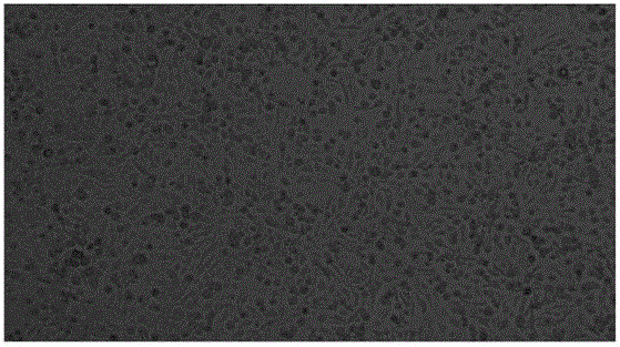
Image


Examples
Embodiment 1
[0037] First, take a fresh placenta from a cesarean delivery woman who has been approved by the family and has negative serological reactions such as HIV, hepatitis A, hepatitis B, and syphilis. 2 PO 4 , 0.132g of Na 2 HPO 4 .12H 2 O, 8g of NaCl, 0.35g of NaHCO 3, 1.0g of D-glucose, 0.10g of streptomycin, and 0.06g of penicillin were prepared by distilling the volume to 1L with water. The basic balanced salt solution was washed repeatedly to remove the residual blood on the surface of the placenta; the amniotic membrane was separated from the placenta, And put it into the basic balanced salt solution for repeated rinsing, and repeat twice; cut the clean amniotic membrane tissue into pieces to obtain amniotic membrane tissue pieces with a diameter of 1mm; add 30ml0.2% IV collagenase to the chopped tissue pieces (gibco, batch number: 1177238), and incubated at 37°C for 2 hours; then centrifuged the tissue block mixed with collagenase at 200g for 5 minutes, and sucked out the...
Embodiment 2
[0039] First, take a fresh placenta from a cesarean delivery woman who has been approved by the family members and whose serological reactions such as HIV, hepatitis A, hepatitis B, and syphilis are negative, and rinses it repeatedly with PBS solution to remove the residual blood on the surface of the placenta; separate the amniotic membrane from the placenta , and put it into PBS to rinse repeatedly, and repeat twice; cut the clean amnion tissue into pieces to obtain amnion tissue pieces with a diameter of 1 mm; add 30ml of 0.2% IV collagenase (gibco , batch number: 1177238), and incubated at 37°C for 2 hours; then centrifuged the tissue block mixed with collagenase at 200g for 5 minutes, and aspirated the collagenase digestion solution; then added 15ml of 0.25% trypsin (sigma, batch number : 110M7362V) / 0.02% EDTA digestion solution, after shaking and digesting at 37°C for 20 minutes, stop digestion with 30ml standard medium containing 10% fetal bovine serum (Hyclone, batch nu...
Embodiment 3
[0041] First, take a fresh placenta from a cesarean delivery woman who has been approved by the family and has negative serological reactions such as HIV, hepatitis A, hepatitis B, and syphilis. 2 PO 4 , 0.132g of Na 2 HPO 4 .12H 2 O, 8g of NaCl, 0.35g of NaHCO 3 , 1.0g of D-glucose, 0.20g of streptomycin, and 0.12g of penicillin were prepared by distilling the volume to 1L with water and washing with basic balanced salt repeatedly to remove the residual blood on the surface of the placenta; separate the amniotic membrane from the placenta, and Put it into the basic balanced salt solution and rinse repeatedly, and repeat twice; cut the clean amnion tissue into pieces to obtain amniotic tissue pieces with a diameter of 1mm; add 30ml0.2% IV collagenase ( gibco, batch number: 1177238), and incubated at 37°C for 2 hours; then centrifuged the tissue block mixed with collagenase at 200g for 5 minutes, and sucked out the collagenase digestion solution; then added 15ml of 0.25% tr...
PUM
| Property | Measurement | Unit |
|---|---|---|
| diameter | aaaaa | aaaaa |
Abstract
Description
Claims
Application Information