
Kit for detecting content of alpha-L-fucosidase

A technology of fucosidase and detection kit, which is applied in the determination/inspection of microorganisms, biochemical equipment and methods, etc., and can solve problems such as necrosis, increased AFU activity, and liver cell degeneration, and achieve low detection limits and improved Active, high-precision effects

Inactive Publication Date: 2015-03-04
CHONGQING QIANDE BIOTECH
View PDF6 Cites 3 Cited by
  • Summary
  • Abstract
  • Description
  • Claims
  • Application Information

AI Technical Summary

Problems solved by technology

At the same time, some studies have reported that the content of AFU in malignant pleural effusions is higher than that in benign pleural effusions, so AFU is a reliable basis for judging benign and malignant pleural effusions; the mechanism is that after the human body is infected with hepatitis B virus (HBV), HBV actively replicates and activates the liver. Tissue inflammatory response factors lead to degeneration and necrosis of liver cells, and then liver cell regeneration and fibrous tissue proliferation, resulting in increased AFU activity

Method used


Image

  • Kit for detecting content of alpha-L-fucosidase
  • Kit for detecting content of alpha-L-fucosidase
  • Kit for detecting content of alpha-L-fucosidase

Examples


Embodiment 1

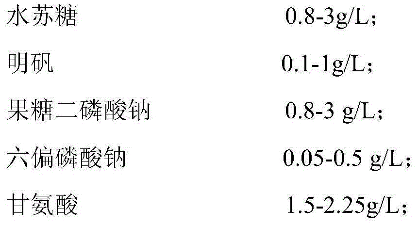
[0043] The kit of the present invention is exemplified as a double reagent, wherein:

[0044] Reagent R1:

[0045]

[0046] Reagent R2:

[0047]

[0048]

Embodiment 2

[0049] Embodiment 2: Kit usage method.

[0050] Reagent R1:

[0051]

[0052] Reagent R2:

[0053]

[0054] 2. Parameter setting of automatic biochemical analyzer:

[0055] (a) Detection wavelength: the main wavelength is 340nm, and the secondary wavelength is 405nm;

[0056] (b) Detection temperature: 37°C;

[0057] (c) Reaction time: 10 minutes, among which, the incubation time is 5 minutes, and the average absorbance change rate △A / min within 3 minutes is measured 2 minutes after adding reagent R2;

[0058] (d) Reaction direction: negative direction.

[0059] 3. Detection steps

[0060] (a) Mix 240ul reagent R1 with 6ul serum sample (to avoid hemolysis);

[0061] (b) Incubate the mixed solution at 37°C for 5 minutes;

[0062] (c) Add 60ul of reagent R2, react for 2 minutes and measure the average absorbance change rate ΔA / min within 3 minutes.

[0063] 4. Calculate the activity of α-L-fucosidase by the average absorbance change rate ΔA / min.

[0064] Following...

Embodiment 3

[0073] Preparation and use of the comparison kit:

[0074] The biological buffer in reagent R1 is Tris buffer 50.0mmol / L, pH 7.0-7.5, and other reagents and experimental methods are the same as in Examples 1 and 2.

[0075]

[0076]

[0077] Reagent R2:

[0078]

[0079] The α-L-fucosidase detection kit prepared in Example 3 was tested for performance, and the method was the same as in Example 2.

[0080] 1) Analytical sensitivity: take a fixed-value α-L-fucosidase sample with a concentration between 2 and 1200U / L to measure the change in absorbance, and repeat the measurement twice to get the average value. The results showed that its analytical sensitivity was 0.5581mA·L / U.

[0081] 2) Minimum detection limit: 5% BSA saline solution is used as a blank sample, and the blank sample should not contain the analyte. The detection was repeated 20 times continuously on the biochemical analyzer, and the detection results were recorded. The results showed that the lowest...


PUM

PropertyMeasurementUnit
wavelengthaaaaaaaaaa

Abstract

The invention discloses a kit for detecting alpha-L-fucosidase. The kit comprises a reagent R1 and a reagent R2 which are independent, wherein the reagent R1 comprises the following components: a bological buffer 1, a metal ion complex, an alpha-L-fucosidase reaction substrate, a surfactant 1 and a preservative 1; and the reagent R2 comprises the following components: a bological buffer 2, 2-chloro-p-nitrophenol-alpha-L-fucosidase, an activating agent, a surfactant 2 and a preservative 2. The kit adopts a dual-reagent mode and has the advantages of high detection sensitivity, low detection limit, wide linear measurement range, high accuracy, good reproducibility, good stability and strong interference resistance. The kit can be used for semi-automatic, full-automatic biochemical analyzer and required detection instrument (biochemical analyzer) commonly used in major hospitals and test centers and is suitable for clinical popularized application, and the rapid diagnosis for emergency is especially achieved.

Description

technical field [0001] The invention relates to the field of biological detection, in particular to an α-L-fucosidase detection kit and a preparation method thereof. Background technique [0002] AFU is an acidic hydrolase, which is widely distributed in the lysosomes of mammalian body fluids and tissue cells, with higher activity in tissues such as liver and kidney. Its main physiological function is to participate in the catabolism of various glycolipids, glycoproteins, mucopolysaccharides and other macromolecular substances containing fucosyl groups. It is synthesized in cells, and its content in the body is always kept at a low level under normal circumstances. The chemical essence of AFU is a glycoprotein, and its relative molecular mass distribution in the body is also different. The relative molecular mass in blood is 2.7×10⁵-3.9×10⁵Dα, which is much larger than that in white blood cells and liver cells. AFU is polymorphic in the human body. When the pH value is 3.8...

Claims


Application Information

Patent Timeline
Patent Type & Authority Applications(China)
IPC IPC(8): C12Q1/34
Inventor 吉权
Owner CHONGQING QIANDE BIOTECH
Learn More