Nano magnetic microspheric ultrasound imaging contrast medium

An ultrasonic imaging and nano-magnetic technology, applied in the medical field, can solve problems such as difficult in vivo imaging, large size of small balls, difficult connection, etc., and achieve good biocompatibility, high mechanical stability, and stable structure

Inactive Publication Date: 2015-03-18
CHENGDU LVKE HUATONG TECH
View PDF1 Cites 1 Cited by
  • Summary
  • Abstract
  • Description
  • Claims
  • Application Information

AI Technical Summary

Problems solved by technology

However, this method still has certain limitations, such as hollow SiO 2 The size of the spheres is too large and does not reach the nanoscale structure, which makes it difficult to be used for in vivo imaging, and it is difficult to carry out further connection without functional group modification
Therefore, the contrast agents currently used in clinical practice cannot simultaneously meet the requirements of good biocompatibility, mechanical stability, targeting performance, nanostructure, and functional group modification.

Method used


Image

  • Nano magnetic microspheric ultrasound imaging contrast medium
  • Nano magnetic microspheric ultrasound imaging contrast medium
  • Nano magnetic microspheric ultrasound imaging contrast medium

Examples


Embodiment 1

[0043] ① Preparation of core / shell polymer nanospheres

[0044]Step 1: First, monodisperse (PE-PAA) copolymer microspheres are prepared by "self-dispersion" polymerization method. Add 0.2mol each of ethylene and acrylic acid, 0.98g AIBN (azobisisobutylcyanide) and 280 mL butyl acetate to a 500mL three-neck round-bottomed reaction flask, and react at a constant temperature of 70°C for 12 hours under the protection of nitrogen. Dry in an oven to obtain monodisperse (PE-PAA) copolymer microspheres.

[0045] Step 2: Prepare core / shell polymer microspheres by seed "self-dispersion" polymerization. Add 3.65 g of (PE-PAA) copolymer microspheres, 0.038 mol each of acrylic acid and divinylbenzene, 0.09 g of AIBN, 100 mL of butyl acetate, and 35 mL of n-hexane into the reaction flask, and keep the temperature at 75 °C for 13 hours to obtain double-layer core / shell polymer nanospheres.

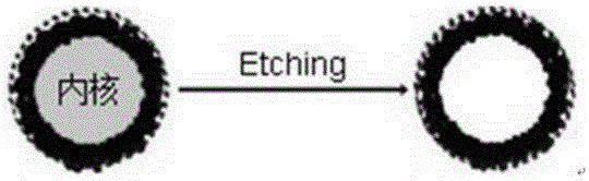
[0046] ② Silica aggregates on the surface of microspheres to form a shell structure

[0047] Usin...


PUM


Abstract

The invention discloses a nano magnetic microspheric ultrasound imaging contrast medium. A microsphere is in a hollow inorganic / organic / inorganic three-layer structure. A hollow cavity is formed in the center of the microsphere with air therein. A Fe3O4 magnetic particle is formed on an external layer of the hollow cavity. A middle layer is a core / shell polymer nano-microsphere which is formed from a polyethylene-polyacrylic acid random copolymer and polyacrylic acid and divinyl benzene through free radical polymerization. A coordinate bond or an ionic bond can be formed between the copolymer and the Fe3O4 magnetic particle, in the internal layer, through carboxyl groups and ferric salts on an internal surface so that the structure is stable. An external layer is silicon dioxide which is connected to the nano-microsphere through a hydrogen bond effect due to the carboxyl groups on the nano-microsphere and silicon hydroxyl groups on the surface of the silicon dioxide. The system is advantaged in that the polymer microsphere belongs to nano-scale particles, wherein the Fe3O4 magnetic particle in the internal layer allows targeted positioning of the contrast medium, the silicon dioxide hollow microsphere is good in biocompatibility and mechanical-stability, and the hollow cavity can generate a strong echo signal under a radiography mode.

Description

technical field [0001] The invention relates to a contrast agent for medical detection, in particular to a multilayer organic / inorganic composite structure nano microsphere with magnetic function, which belongs to the field of medical technology. Background technique [0002] In recent years, molecular imaging technology has attracted extensive attention because it can provide important information for the diagnosis and monitoring of diseases in living tissues (Campbell R. E., Chang C. J. Curr Opin Chem Biol , 2010, 14(1):1-2.). Among many imaging modes, ultrasound imaging, as a non-invasive biomedical imaging mode, has important research value due to its advantages of safe and timely imaging and diagnosis, low price, lightness and quickness, and wide application. However, the difference in echo signals between different biological tissues often hinders the accuracy of diagnosis. In order to better obtain accurate information of specific tissues, people usually use the meth...

Claims


Application Information

Patent Timeline
IPC IPC(8): A61K49/22
Inventor 不公告发明人
Owner CHENGDU LVKE HUATONG TECH
Why Eureka
Try Eureka