Preparation and application of anti-Coxsackie virus A16 monoclonal antibody

A coxsackie virus, A16 technology, applied in the field of biotechnology and immunology, can solve the problem of no monoclonal antibody

Active Publication Date: 2015-04-15
INST PASTEUR OF SHANGHAI CHINESE ACADEMY OF SCI
View PDF3 Cites 5 Cited by
  • Summary
  • Abstract
  • Description
  • Claims
  • Application Information

AI Technical Summary

Problems solved by technology

However, there is no monoclonal antibody against CA16 in t

Method used


Image

  • Preparation and application of anti-Coxsackie virus A16 monoclonal antibody
  • Preparation and application of anti-Coxsackie virus A16 monoclonal antibody
  • Preparation and application of anti-Coxsackie virus A16 monoclonal antibody

Examples


preparation example Construction

[0099] For the preparation of CA16 / SZ05 inactivated virus, see Cai Y et al., Vaccine2013Apr26; 31(18):2215-21. After mixing 4ug of inactivated virus (50ul volume) with an equal volume of aluminum adjuvant and 25ug CpG adjuvant, female Balb / c mice were immunized intraperitoneally for 6 weeks, and immunized once at 0 weeks, 2 weeks, 4 weeks, and 6 weeks. At week 7, mouse serum was taken to detect the neutralization titer. One mouse with the highest neutralizing titer was boosted with 10ug of inactivated CA16 virus through the tail vein at the 8th week. After 3 days, the mouse spleen was taken for the preparation of hybridoma cells.

[0100] Preparation and screening of hybridoma cell lines

[0101] Three days after the tail vein booster immunization of the mice, the spleen cells of the mice were fused with myeloma cells SP2 / 0 through PEG1500 to prepare hybridoma cells. After 9 days, neutralizing antibodies specifically secreted against CA16 were screened by ELISA and microneu...

Embodiment 1

[0128] Embodiment 1, establishment of hybridoma cell line 9B5

[0129] Hybridoma cells were obtained by fusion of splenocytes from mice immunized with inactivated CA16 and myeloma cell SP2 / 0. The supernatant of hybridoma cells was analyzed by ELISA and microneutralization titration experiments, and a hybridoma cell line was screened out. The antibody secreted by it could bind CA16 virus and had neutralizing effect in vitro. The hybridoma cell line was named 9B5.

Embodiment 2

[0130] Embodiment 2, characteristic analysis of monoclonal antibody 9B5

[0131] The characteristics of monoclonal antibody secreted by hybridoma cell line 9B5 were analyzed. Subtype identification showed that the light chain of monoclonal antibody 9B5 belonged to kappa chain, and the heavy chain belonged to IgG2B.

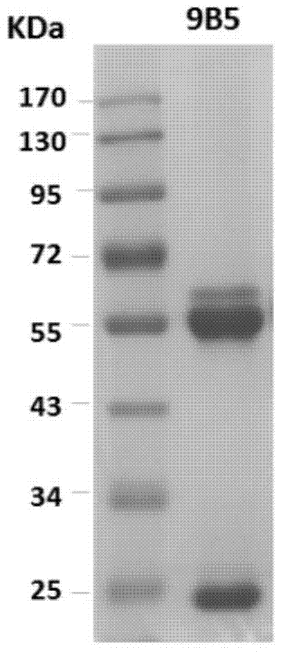
[0132] SDS-PAGE analysis showed that the heavy chain and light chain of mAb 9B5 were about 50KD and 25KD respectively, as shown in figure 1 .

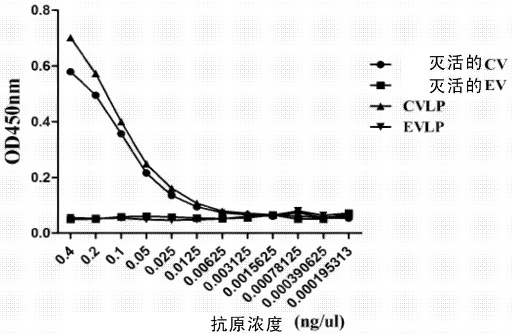
[0133] The reactivity of monoclonal antibody 9B5 with different antigens was detected by ELISA, and the results showed that 9B5 reacted with inactivated CA16 virus, but not with inactivated EV71 virus, indicating that the antibody specifically recognized CA16. In addition, none of the monoclonal antibodies reacted with the CA16 capsid protein VPO, VP1, or VP3 recombinantly expressed in E. coli, suggesting that the monoclonal antibody recognized a conformational epitope.


PUM


Abstract

The invention relates to preparation and application of an anti-Coxsackie virus A16 (CA16) monoclonal antibody. The invention reveals a mouse source anti-CA16 monoclonal antibody, which has good binding specificity, can be used for sensitive detection of Coxsackie A16 virus, and has strong anti-virus infection capacity.

Description

technical field [0001] The invention belongs to the field of biotechnology and immunology; more specifically, the invention relates to the preparation and application of an anti-Coxsackie virus A16 monoclonal antibody. Background technique [0002] Coxsackievirus A16 (Coxsackievirus A16, CA16) belongs to the Picornaviridae Enterovirus genus, and CA16 is one of the main pathogens of hand, foot and mouth disease (HFMD), and most patients infected by CA16 The clinical symptoms are relatively mild, and most of them can heal by themselves, including fever, oral ulcers, and red herpes on the hands and feet. But more and more evidence shows that CA16 infection can also cause serious diseases such as nervous system damage, and even lead to death. There is currently no antiviral drug for the treatment of CA16 infection, and a vaccine is still under development. This is partly due to the lack of reagents available for the qualitative and quantitative analysis of CA16, such as CA16-s...

Claims


Application Information

Patent Timeline
IPC IPC(8): C07K16/10C12N15/13C12N15/63C12N5/10C12N1/15C12N1/19C12N1/21A61K39/42A61P31/14G01N33/577
Inventor 黄忠石金平刘庆伟
Owner INST PASTEUR OF SHANGHAI CHINESE ACADEMY OF SCI
Why Eureka
Try Eureka