Method for preparing microfiber hemostatic collagen sponge

A technology of collagen sponge and microfiber, which is applied in medical science, absorbent pads, bandages, etc., can solve problems such as difficulty in maintaining the inherent shape, poor mechanical properties, and easy degradation, and achieve good hemostatic performance, efficient water absorption, and simple preparation process Effect

Inactive Publication Date: 2015-04-29
无锡贝迪生物工程股份有限公司
View PDF2 Cites 17 Cited by
  • Summary
  • Abstract
  • Description
  • Claims
  • Application Information

AI Technical Summary

Problems solved by technology

[0004] The purpose of the present invention is to provide a method for preparing microfiber hemostatic collagen sponge, the prepared microfiber hemostatic collagen sponge has uniform porous structure, good hydrophilicity, high mechanical strength and degradation resistance and good hemostatic performance, to solve The traditional collagen sponge is easy to degrade, has poor mechanical properties, is easy to collapse during application, and is difficult to maintain its inherent shape.

Method used


Image

  • Method for preparing microfiber hemostatic collagen sponge
  • Method for preparing microfiber hemostatic collagen sponge
  • Method for preparing microfiber hemostatic collagen sponge

Examples


Embodiment 1

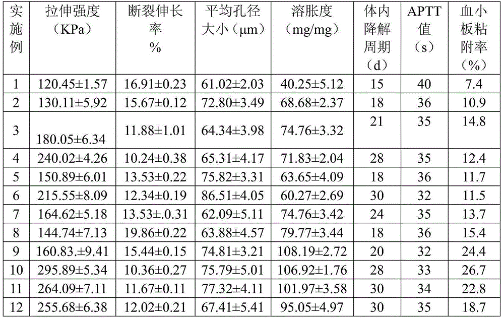
[0026] At 4°C, mix 0.5 mg / mL collagen acetate solution with 0.085M phosphate buffer at a volume ratio of 16:1, adjust pH to 5 with 0.1M NaOH solution, stir and mix well, and transfer to a 15°C water bath After 2 h, the solidified assembled collagen solution was centrifuged at 4000 rmp / min for 10 min, the supernatant was discarded, and the salt was removed by dialysis for 1 d. The assembled collagen solution was freeze-dried to make a sponge, which was cross-linked in a vacuum drying oven at 80°C for 24 hours to obtain a microfiber hemostatic collagen sponge. The properties of the obtained sponge are shown in Table 1.

Embodiment 2

[0028] At 4°C, mix 1 mg / ml collagen acetic acid solution with 0.09M phosphate buffer at a volume of 8:1, adjust pH to 6 with 0.1M NaOH solution, stir and mix well, and then transfer to a 20°C water bath. After 4 hours, the solidified collagen solution was centrifuged at 5000 rmp / min for 10 minutes, the supernatant was discarded, and the salt was removed by dialysis for 2 days. The assembled collagen solution was freeze-dried to form a sponge, and cross-linked in a vacuum drying oven at 100°C for 24 hours to obtain a microfiber hemostatic collagen sponge. The properties of the obtained sponge are shown in Table 1.

Embodiment 3

[0030] At 4°C, mix 2 mg / ml collagen acetic acid solution with 0.075M phosphate buffer at a volume of 4:1, adjust pH to 7 with 0.1M NaOH solution, stir and mix well and transfer to a 25°C water bath. After 6 h, the solidified assembled collagen solution was centrifuged at 6000 rmp / min for 10 min, the supernatant was discarded, and the salt was removed by dialysis for 3 d. The assembled collagen solution was freeze-dried to make a sponge, and cross-linked in a vacuum drying oven at 110° C. for 24 hours to obtain a microfiber hemostatic collagen sponge. The properties of the obtained sponge are shown in Table 1.


PUM

PropertyMeasurementUnit
concentrationaaaaaaaaaa

Abstract

The invention relates to a method for preparing microfiber hemostatic collagen sponge. The method comprises the following steps of: aggregating collagen molecules by the charge effect by adjusting pH, temperature and the salt concentration of collagen protein solution, freeze-drying the obtained and assembled collagen, then preparing into the collagen protein sponge, and then carrying out thermal crosslinking to obtain the microfiber hemostatic collagen sponge. The microfiber hemostatic collagen sponge prepared by adopting the method has uniform porous structure, good hydrophilic performance, high mechanical strength and degradation resistance and good hemostatic performance, and the defects that the traditional collagen sponge is easy in degradation, poor in mechanical property, easy in collapse in the application process and difficult in maintenance of the inherent form and the like are solved. The preparation method is simple and feasible, and is stable in process, and the prepared microfiber hemostatic collagen sponge can be widely applied in hemostasis and restoration of wounds of burns, traumas and surgical operation, and has good application prospect.

Description

technical field [0001] The invention relates to a method for preparing a microfiber hemostatic collagen sponge, which belongs to the technical field of biomedical materials. Background technique [0002] Biomedical materials are a class of high-tech new materials developed in the past 30 years. Among them, absorbable hemostatic materials have gradually attracted the attention of the medical community with the increase of traffic accidents and major disasters. With the rapid development of modern science and technology, the research on hemostatic materials has made very rapid progress, and various new hemostatic materials are constantly emerging, and their performance is getting better and better. Currently commonly used hemostatic materials include absorbable fibrin glue, chitosan, absorbable gelatin sponge, cyanoacrylate, oxidized cellulose and oxidized regenerated cellulose. Biomedical hemostatic materials with definite hemostatic effect, good biocompatibility and control...

Claims


Application Information

Patent Timeline
Patent Type & Authority Applications(China)
IPC IPC(8): C08J9/28C08J3/24C08L89/00A61L15/32A61L15/42A61L15/64
Inventor 任伟业赵颖陆金婷林芳
Owner 无锡贝迪生物工程股份有限公司
Why Eureka
Try Eureka