Separation method of limbal stem cells

A corneal limbal stem cell and separation method technology, applied in the field of corneal limbal stem cell separation, can solve the problems of fibroblasts being difficult to adhere to the wall, reducing cell purity, etc., and achieving the effects of reducing damage, increasing cell yield, and improving purity

Inactive Publication Date: 2016-01-06
GUANGZHOU SALIAI STEMCELL SCI & TECH CO LTD
View PDF2 Cites 9 Cited by
  • Summary
  • Abstract
  • Description
  • Claims
  • Application Information

AI Technical Summary

Problems solved by technology

[0007] Although the enzymatic digestion method in the existing primary isolation method of limbal stem cells can obtain a large number of cells in a short period of time, trypsin will affect the adhesion of the cells and cause fibrogenesis in the later isolation process. The cells are not easy to adher

Method used


Image

  • Separation method of limbal stem cells
  • Separation method of limbal stem cells
  • Separation method of limbal stem cells

Examples


Embodiment 1

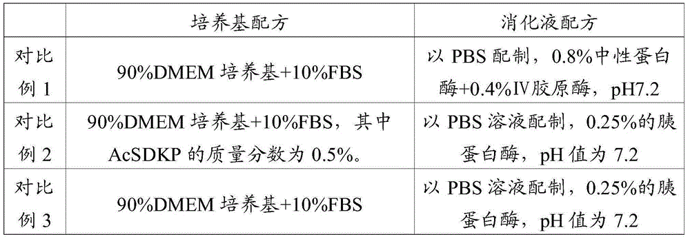
[0053] Compound enzyme solution: prepared in PBS, 0.75% neutral protease + 0.50% IV collagenase, pH7.2

[0054] Medium: 90% DMEM medium + 10% FBS, wherein the mass fraction of AcSDKP is 0.5%.

[0055] The eyeballs of dead fetuses with a gestational age of 3-7 months were soaked in PBS containing double-antibody (penicillin-streptomycin, 1×) for 3 minutes in a sterile environment, and then rinsed with PBS without double-antibody.

[0056] Under the operating microscope, use tissue forceps and corneal scissors to cut the human corneal limbal tissue, and then use scissors to cut the tissue into about 1mm 3 blocky in size.

[0057] According to the volume of the tissue block, every 1cm 3 Add 5ml compound enzyme solution to the tissue block, digest at 37°C for 15min, add PBS containing 10% FBS to stop digestion per 1ml of digestion solution, filter with 100um mesh, centrifuge at 1000rpm for 5min, discard the supernatant, add culture medium, dilute at 1×10 5 The density of cell / m...

Embodiment 2

[0060] Compound enzyme solution: prepared in PBS, 0.8% neutral protease + 0.4% IV collagenase, pH7.2

[0061] Medium: 90% DMEM medium + 10% FBS, wherein the mass fraction of AcSDKP is 0.8%.

[0062] The eyeballs of dead fetuses with a gestational age of 3-7 months were soaked in PBS containing double-antibody (penicillin-streptomycin, 1×) for 3 minutes in a sterile environment, and then rinsed with PBS without double-antibody.

[0063] Under the operating microscope, use tissue forceps and corneal scissors to cut the human corneal limbal tissue, and then use scissors to cut the tissue into about 1mm 3 blocky in size.

[0064] According to the volume of the tissue block, every 1cm 3 Add 5ml compound enzyme solution to the tissue block, digest at 37°C for 10min, add PBS containing 10% FBS to each 1ml of digestion solution to stop digestion, filter with 100um mesh, centrifuge at 1000rpm for 5min, discard the supernatant, add medium, dilute at 1×10 5 The density of cell / ml was ...

Embodiment 3

[0067] Compound enzyme solution: prepared in PBS, 0.78% neutral protease + 0.45% IV collagenase, pH7.2

[0068] Medium: 90% DMEM medium + 10% FBS, wherein the mass fraction of AcSDKP is 1%.

[0069] The eyeballs of dead fetuses with a gestational age of 3-7 months were soaked in PBS containing double-antibody (penicillin-streptomycin, 1×) for 3 minutes in a sterile environment, and then rinsed with PBS without double-antibody.

[0070] Under the operating microscope, use tissue forceps and corneal scissors to cut the human corneal limbal tissue, and then use scissors to cut the tissue into about 1mm 3 blocky in size.

[0071] According to the volume of the tissue block, every 1cm 3 Add 5ml compound enzyme solution to the tissue block, digest at 37°C for 20min, add PBS containing 10% FBS to each 1ml digestion solution to stop digestion, filter with a 100um mesh sieve, centrifuge at 1000rpm for 5min, discard the supernatant, add the culture medium, and dilute with 1×10 5 The ...


PUM


Abstract

The invention relates to the field of stem cell culture and in particular relates to a separation method of limbal stem cells. The separation method has the beneficial effects that limbal tissues are subjected to enzymolysis under the combined action of a compound enzyme by combining neutral protease with collagenase IV, thus ensuring obtainment of the cells through enzymolysis in a short time, reducing damages to the cells, increasing the cell yield and improving the purity of the obtained cells; the method is simple and convenient to operate, dispenses with high-end instruments and equipment and causes smaller damages to the cells; experiments show that by adopting the method provided by the invention to separate the limbal stem cells, every 1cm<3> of limbal tissues can obtain 8.4*10<5>-9.0*10<5> limbal stem cells, the positive rate of an antibody CX43 in the cells is 0, the positive rate of AE5 is 0.1%, the positive rate of BrdU is 70.1%, and the positive rate of PCNA is 82.1%; the separation method has obviously better effects than the prior art.

Description

technical field [0001] The invention relates to the field of stem cell culture, in particular to a method for separating corneal limbal stem cells. Background technique [0002] The corneal limbus is the junction of the cornea, conjunctiva and sclera. The distinguishing mark from the cornea is the termination of Bowman's membrane; the distinguishing mark from the conjunctiva is that it does not contain goblet cells and is about 1 mm wide. There are only epithelial and stroma layers here. The epithelial cell layer is more than 10 layers, arranged irregularly, the cells are small cylindrical, and the nucleus is deeply stained. The deep basal cells are a layer of small cylindrical or cuboid cells, and the nuclei are oval and parallel to the surface. In the basal The papillae are formed in the papilla, forming a special "palisade"-like epithelial structure, which contains pigment and rich vascular network, and is closely connected with the basement membrane. [0003] Limbal ste...

Claims


Application Information

Patent Timeline
IPC IPC(8): C12N5/074
Inventor 陈海佳王一飞葛啸虎吴子杰张维敏
Owner GUANGZHOU SALIAI STEMCELL SCI & TECH CO LTD
Why Eureka
Try Eureka