Protein chip for typing detection on helicobacter pylori infection

A Helicobacter pylori and protein chip technology, applied in measurement devices, analytical materials, biological tests, etc., can solve the problems of inability to judge type I and type II infections, limited positive rate, etc.

Inactive Publication Date: 2016-01-27
TAIZHOU SYNO GENE DIGITAL TECH CO LTD
View PDF2 Cites 5 Cited by
  • Summary
  • Abstract
  • Description
  • Claims
  • Application Information

AI Technical Summary

Problems solved by technology

The detection antigen used in the above products may be a single Ure protein (such as the gold standard method), which can only detect the anti-Ure antibody in the patient's serum, so the positive rate is limited; Judgment is type I and type II infection

Method used


Image

  • Protein chip for typing detection on helicobacter pylori infection
  • Protein chip for typing detection on helicobacter pylori infection

Examples


Embodiment 1

[0018] Example 1 Preparation of protein chip

[0019] Specifically include the following steps:

[0020] 1. Preparation of NC Membranes

[0021] (1) Soak the NC membrane with a pore size of 0.45 μm in distilled water first, take it out and dry it, soak it in 0.05mol / L carbonate buffer solution and then dry it in air.

[0022] (2) Cut the NC film into square pieces of 0.9 cm×0.9 cm with a semi-automatic film cutting machine, and set aside.

[0023] (3) Take the diluted 0.5mg / ml CagA antigen, 1.0mg / ml VacA antigen, and 1.0mg / ml Ure antigen and put them into the corresponding sample boxes in the spotting instrument respectively.

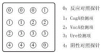
[0024] (4) Under the condition of temperature control (≤10°C), place the nitrocellulose membrane flatly in the spotting tank, and use the MicroGrid-2 spotting instrument to place the antigen solution, positioning solution, and quality control control solution in the form of a dot matrix. Form points on the nitrocellulose membrane, every 3 points as a...

Embodiment 2

[0034] Example 2 Preparation of a protein chip kit for typing and detecting Helicobacter pylori infection

[0035] The kit is composed of washing solution, chromogen and the protein chip of Example 1.

[0036] 1. Prepare washing solution

[0037] (1) Measure 500ml TBE buffer solution with a 1000ml graduated cylinder and pour it into a 2000ml beaker;

[0038] (2) Measure 100ml of saturated ammonium sulfate solution with a 100ml graduated cylinder, and pour it into a 2000ml beaker;

[0039] (3) Turn on the magnetic stirrer switch, adjust the rotating speed until the liquid can be fully stirred evenly;

[0040] (4) Measure 20ml Tween-20 with a 100ml measuring cylinder, pour it into a 2000ml beaker, and continue stirring;

[0041] (5) Measure 500ml deionized water with a 500ml graduated cylinder;

[0042] (6) Pour about 100ml into a measuring cylinder filled with Tween-20, shake the measuring cylinder to clean the residual Tween-20, and then pour into a 2000ml beaker;

[0043...

Embodiment 3

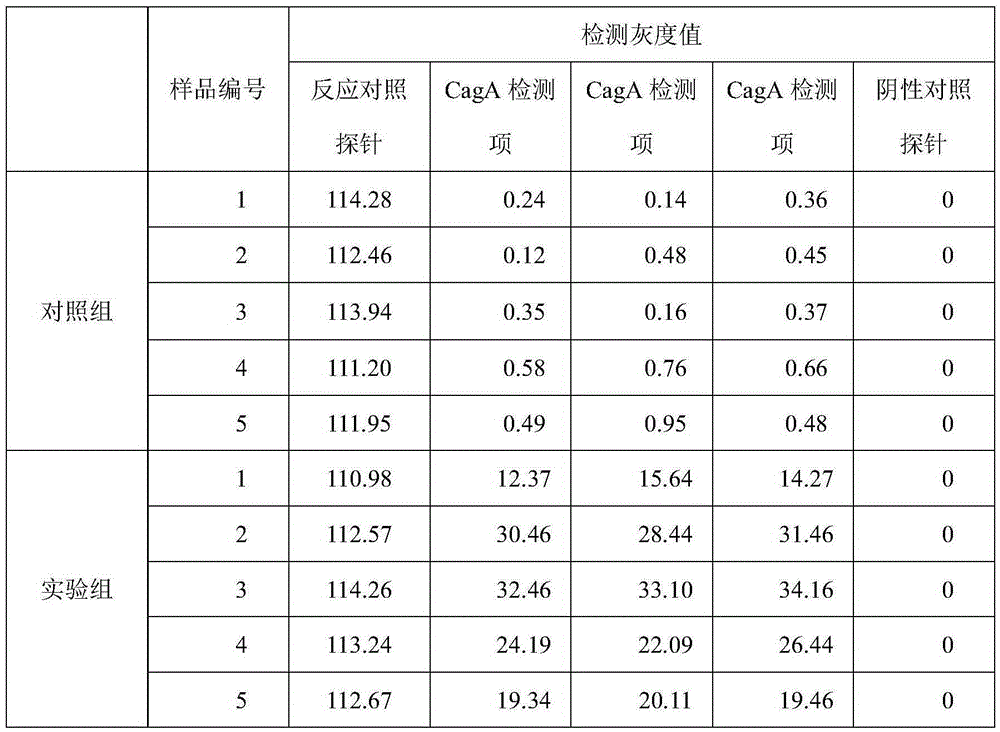
[0054] Example 3 Detection Verification

[0055] Five samples of serum from normal people and patients infected with Helicobacter pylori were collected and divided into control group and experimental group. Using the kit of Example 2, strictly follow the steps below to compare and verify the detection results of the protein chip of the present invention.

[0056] Detection steps:

[0057] 1) Moisturizing membrane: Take 160 μl (4 drops) of washing solution and add it to the membrane surface of the detection window of the chip to evenly wet the membrane surface;

[0058] 2) Adding samples: Take 200 μl of the processed serum to be tested and add it to the membrane surface of the detection window of the chip;

[0059] 3) Washing: After the sample is fully infiltrated, in order to ensure the washing effect, wash in two steps: take 120 μl (3 drops) of washing solution, add it to the membrane surface of the chip detection window, and after the washing solution penetrates, take anot...


PUM


Abstract

The invention provides a protein chip for typing detection on helicobacter pylori infection. The protein chip comprises a basement membrane and antigens which are respectively dotted on the basement membrane in the form of dot matrix, wherein the antigens include three types, i.e. CagA (cytotoxin-associated gene) antigens, VacA (Vacuolating cytotoxin A) antigens and Ure (ureaplasma urealyticum) antigens. Due to the fact that the detection antigens used by the protein chip are three single proteins, i.e. Ure, CagA and VacA, specific IgG antibodies for the three antigens in the blood serum of a patient can be detected at the same time, I-type infection or II-type infection can be judged, and clinic treatment is better facilitated; furthermore, the protein chip has the advantages of high detection sensitivity, quick, simple and convenient detection, instant readability, small using amount of antigen and the like.

Description

technical field [0001] The invention relates to a protein chip for typing and detecting Helicobacter pylori infection, belonging to the technical field of biochips. Background technique [0002] Helicobacter pylori (Hp) can grow and multiply in the human body, and can be excreted from the body through feces and saliva. In developing countries, nearly 80% of children under the age of 10 and more than 90% of adults are infected. [0003] After years of research, it has been found that Hp cytotoxin-associated protein A (CagA), cytovacuole toxin A protein (VacA) and urease (Ure) can stimulate the body to produce a strong immune response, which can be detected in the serum of patients infected with Hp to the corresponding antibody. The combined detection of antibodies to these antigens can not only improve the sensitivity and specificity of the detection of Helicobacter pylori, but also obtain more information about the infectious pathogen, which can provide assistance for clin...

Claims


Application Information

Patent Timeline
Patent Type & Authority Applications(China)
IPC IPC(8): G01N33/68G01N33/569
CPCG01N33/68G01N33/56911G01N2333/195
Inventor 闫小君阎平希王小明薛小平曾德隆王艳玲郭松
Owner TAIZHOU SYNO GENE DIGITAL TECH CO LTD
Why Eureka
Try Eureka