
Acid phosphatase staining method for distinguishing malignant serosal cavity effusion mesothelial cells from cancer cells and application thereof

A serosal cavity effusion and acid phosphatase technology, applied in the field of acid phosphatase staining, can solve the problems of difficult cancer cell differentiation, pathologist’s inability to make judgments, and low sensitivity of exfoliated cytology examination, etc. Convenience, reduced workload, guaranteed specificity and sensitivity

Inactive Publication Date: 2016-03-23
HEFEI JINCIKANG BIOTECH CO LTD
View PDF4 Cites 0 Cited by
  • Summary
  • Abstract
  • Description
  • Claims
  • Application Information

AI Technical Summary

Problems solved by technology

Cytological examination makes judgments based on the morphological characteristics of the cells, and the mesothelial cells in the serosal cavity have various shapes due to hyperplasia, degeneration and other reasons, and it is not easy to distinguish them from cancer cells only by Papanicolaou staining. Exfoliative cytology of effusions often has low sensitivity (40%-60%), and inexperienced pathologists cannot even make a judgment

Method used


Image

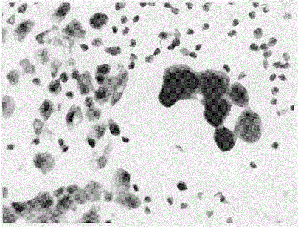
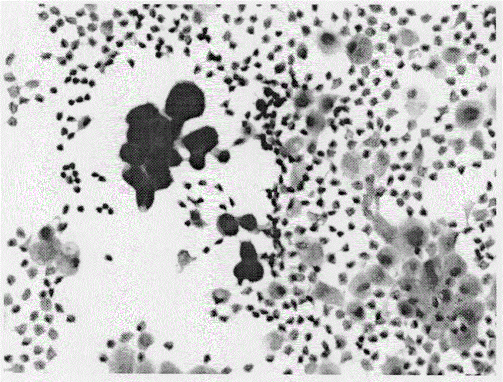
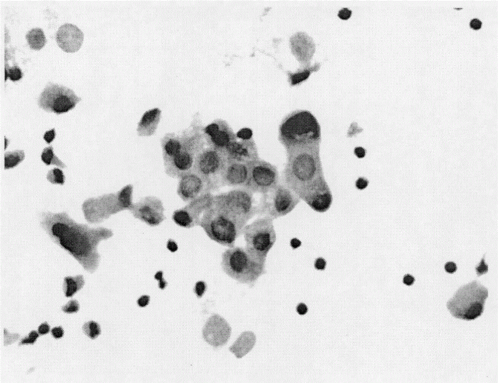
  • Acid phosphatase staining method for distinguishing malignant serosal cavity effusion mesothelial cells from cancer cells and application thereof
  • Acid phosphatase staining method for distinguishing malignant serosal cavity effusion mesothelial cells from cancer cells and application thereof
  • Acid phosphatase staining method for distinguishing malignant serosal cavity effusion mesothelial cells from cancer cells and application thereof

Examples


Embodiment 1

[0012] 1. Acid Phosphatase Staining Kit Components:

[0013] (1) Fixative: 5-10% formalin

[0014] (2) Staining solution: 5-20g / l naphthol AS-BI phosphate dissolved in dimethylformamide

[0015] (3) Fuchsin hydrochloride: 2-10% fuchsin hydrochloride

[0016] (4) Sodium nitrite: 0.1-0.4MNaNO2

[0017] (5) Sodium acetate solution: 2-5M, pH4.5-5.5

[0018] (6) Distilled water

[0019] (7) Nuclear counterstaining solution: 2.5g of hematoxylin, 20ml of absolute alcohol, 5g of potassium aluminum sulfate, 330ml of distilled water, 250mg of sodium iodate, 150ml of glycerol, and 10ml of glacial acetic acid.

[0020] In addition, the above kit may also include an enzyme-labeled liquid-based cytology preservation solution, and its specific composition is as follows:

[0021] (1) 15% Methanol

[0022] (2) 0.1mM EDTA

[0023] (3) 0.1mM NaCl

[0024] (4) 0.01M sodium acetate buffer, pH5.2

[0025] (5) 5% Proclin300

[0026] (6)ddH 2 O.

[0027] 2. Dyeing steps:

[0028] (1) Cel...


PUM


Abstract

The invention relates to the field of auxiliary diagnosis, in particular to an acid phosphatase staining method for distinguishing malignant serosal cavity effusion mesothelial cells from cancer cells, a relevant reagent and the application of the method. By the adoption of the method, erythrocytes in a sample can be removed, and background influence can be eliminated; meanwhile, mesothelial cells and malignant cells can be distinguished distinctly, a doctor is helped to find malignant cells quickly and accurately and make a diagnosis on this basis, and diagnosis accuracy is improved greatly.

Description

technical field [0001] The invention relates to the field of auxiliary diagnosis, in particular to an acid phosphatase staining method for distinguishing malignant serous cavity effusion mesothelial cells from cancer cells, related reagents and applications thereof. Background technique [0002] The serous cavity is the gap between the serous parietal layer and the visceral layer, and is divided into the pleural cavity, peritoneal cavity, and pericardial cavity. There is a little serous fluid in the cavity, which acts as a lubricant, and it can increase in pathological conditions, forming pleural effusion, ascites, pericardial effusion, etc. The serosal effusions submitted for clinical examination are all pathological, and the cellular components in the serosal effusion include mesothelial cells, blood cells of non-epithelial origin, and tumor cells may also exist. Benign lesions (such as pneumonia, pulmonary infarction, influenza, liver abscess, tuberculosis, heart failure...

Claims


Application Information

Patent Timeline
IPC IPC(8): G01N1/30
Inventor 吴广平王阳方圣云方圣文
Owner HEFEI JINCIKANG BIOTECH CO LTD
Learn More