Isolation and primary culture methods of chicken small intestinal epithelial cells

A technology for small intestinal epithelial cells and primary culture, which is applied in cell dissociation methods, cell culture active agents, gastrointestinal cells, etc. Good condition, low mortality rate, good cell growth condition

Pending Publication Date: 2016-04-13
SHANXI AGRI UNIV
View PDF2 Cites 7 Cited by
  • Summary
  • Abstract
  • Description
  • Claims
  • Application Information

AI Technical Summary

Problems solved by technology

[0009] In order to solve the problems that chicken small intestinal epithelial cells are difficult to separate and cultivate in vitro, the existing separation process is cumbersome

Method used


Image

  • Isolation and primary culture methods of chicken small intestinal epithelial cells
  • Isolation and primary culture methods of chicken small intestinal epithelial cells
  • Isolation and primary culture methods of chicken small intestinal epithelial cells

Examples


experiment example 1

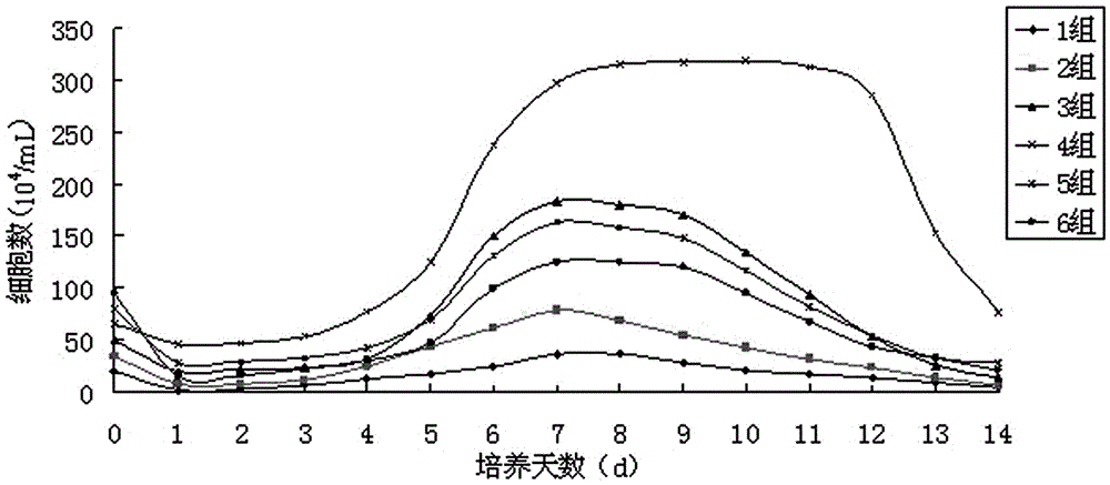
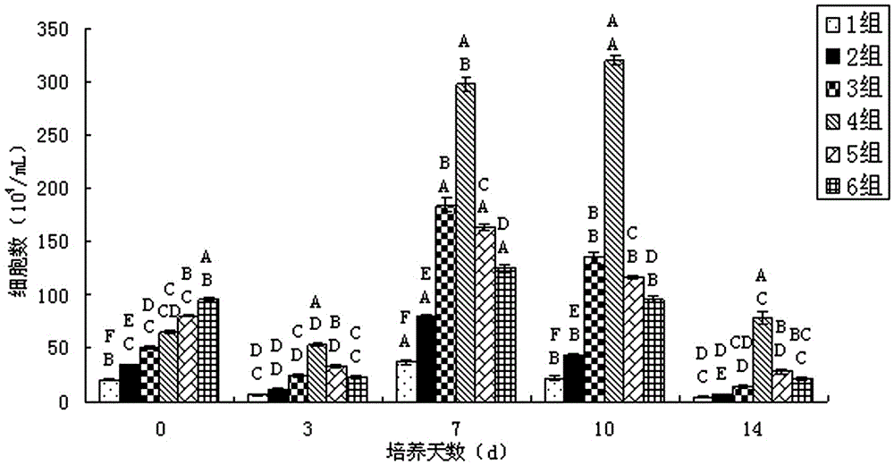
[0031] Experimental Example 1: Screening and Culture of the Optimal Cell Seeding Density of Chicken Small Intestinal Epithelial Cells

[0032] According to the number of cells per ml obtained by cell counting, it is divided into six gradients, as shown in Table 1. The above-mentioned diluted cell mass dilutions were inoculated into 6-well cell culture plates according to the data of different gradients, and each gradient was repeated 3 times. , placed at 40°C, 7%CO 2 Cultivate in the incubator for 36 hours, observe the growth state of the cells, and calculate the cell attachment rate. After 48 hours, the culture medium was replaced, and the growth and adhesion of the cells were observed every day. The number of living cells was counted by trypan blue staining, and the observation was continued for 14 days. The growth curve of chicken small intestinal epithelial cells was made with the culture time of chicken small intestinal cells as the abscissa and the number of viable cell...

experiment example 2

[0041] Experimental Example 2: Screening of the Best Serum for Chicken Small Intestinal Epithelial Cells

[0042]Add different concentrations of fetal bovine serum and chicken serum to the culture medium, observe the growth of the cells, divide the two serums into group A (fetal bovine serum) and group B (chicken serum), and divide the serum into 0% , 5%, 10% different concentrations. Before the adherence difference, both groups A and B were cultured with 10% serum concentration medium to culture enzyme-digested cells, which was helpful for the adherent growth of non-intestinal epithelial cells such as fibroblasts. After poor adherence, culture the purified chicken small intestinal epithelial cells with a culture medium with a serum concentration of 5%, and change the medium every two to three days. Proliferation, cells by approximately 6.5×10 5 / mL inoculated into 6-well culture plates, with 3 replicates in each group, placed at 40°C, 7% CO 2 Culture in the incubator for 1...

experiment example 3

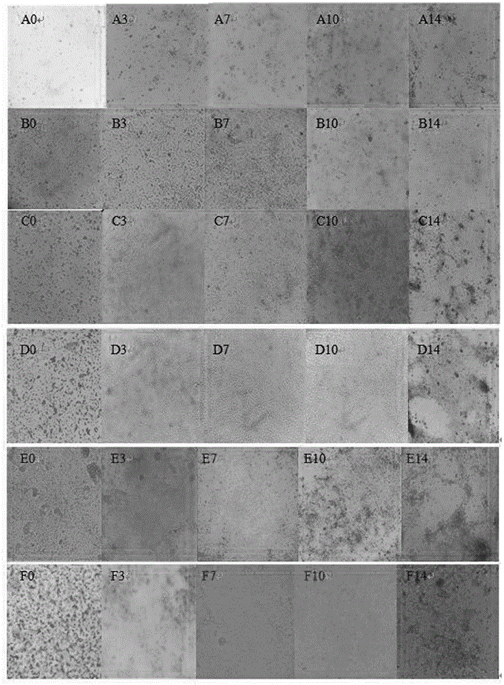
[0049] Experimental Example 3: Identification of Chicken Small Intestinal Epithelial Cells

[0050] (1) Morphological identification of cells: After the primary culture of chicken intestinal epithelial cells started, live cells were directly observed through an inverted phase-contrast microscope, mainly to observe the growth and proliferation of cells attached to the wall, including the changes in cell shape and size at different times.

[0051] Cells that have just been digested from the small intestine of chickens are inoculated into cell culture plates, and the cells are observed under a microscope as Figure 7 As shown, most of them are different in size, round in shape, and transparent, and most of them digested are cell clusters. 2 Within 24 hours of culture, the cells began to adhere to the wall. Most of the newly adhered cells were distributed in the shape of islands, and some of them were scattered in the shape of flat irregular polygons and spindles. Most of the cell...


PUM


Abstract

The invention belongs to the technical field of cell isolation and cultivation. In order to solve the problems that the chicken small intestinal epithelial cells are difficult to isolate and culture in vitro at present, the existing isolation process is troublesome and time-consuming, the obtained cells are low in purity and the like, the invention provides isolation and primary culture methods of the chicken small intestinal epithelial cells. The methods comprise the steps of taking chick embryo small intestines, cleaning the small intestines to remove mesenteries, digesting small intestine tissues by protease to obtain the chicken small intestinal epithelial cells, performing adherent difference culture on the cells, removing adherent parenchyma cells, collecting intestinal epithelial cells, inoculating by taking the inoculum density being about 6.5X10⁵/mL for culturing, the chicken small intestinal epithelial cells obtained by culturing is maximum in quantity, low in death rate, and best in growing status. The primary culture is performed by adopting a culture solution in which chicken serum is added, and the growing status of the obtained cells is better. The chicken small intestinal epithelial cells which are good in growing status, normal in shape, high in activity are successfully cultured, the primary and isolation culture methods of the chicken small intestinal epithelial cells are successfully established, and the technical support is provided for follow-up tests.

Description

technical field [0001] The invention belongs to the technical field of cell separation and culture, and in particular relates to a method for separation and primary culture of chicken small intestinal epithelial cells. Background technique [0002] Intestinal epithelial cells (IEC) play an important role in many aspects of intestinal physiology and pathology. The gut is the largest immune organ and it is important for the mucosal-borne immune response to food antigens. Therefore, the study of intestinal epithelial cells is helpful to explore the physiological and pathological functions of the intestinal tract. Intestinal epithelial cells can act as a barrier to prevent the entry of potentially harmful substances and the migration of underlying cells. In vitro culture experiments have shown that intestinal epithelial cells affect the changes in the number of cells in the microenvironment. [0003] In vitro primary cultures of intestinal epithelial cells are difficult becaus...

Claims


Application Information

Patent Timeline
IPC IPC(8): C12N5/071
CPCC12N5/0679C12N5/0625C12N2500/32C12N2500/84C12N2501/11C12N2501/33C12N2501/91C12N2501/998C12N2509/00
Inventor 张利环李慧锋朱芷葳张瑜李玲香
Owner SHANXI AGRI UNIV
Why Eureka
Try Eureka