
Polymer enzyme-antibody and preparation method thereof

A polymer enzyme and antibody technology, applied in the field of medical detection, can solve the problems of long incubation time, loose overall structure of the secondary antibody, misdiagnosis, etc., and achieve the effect of improving the sensitivity and improving the detection sensitivity.

Active Publication Date: 2016-05-11
XIAMEN TALENT BIOMEDICAL TECH CO LTD
View PDF2 Cites 19 Cited by
  • Summary
  • Abstract
  • Description
  • Claims
  • Application Information

AI Technical Summary

Problems solved by technology

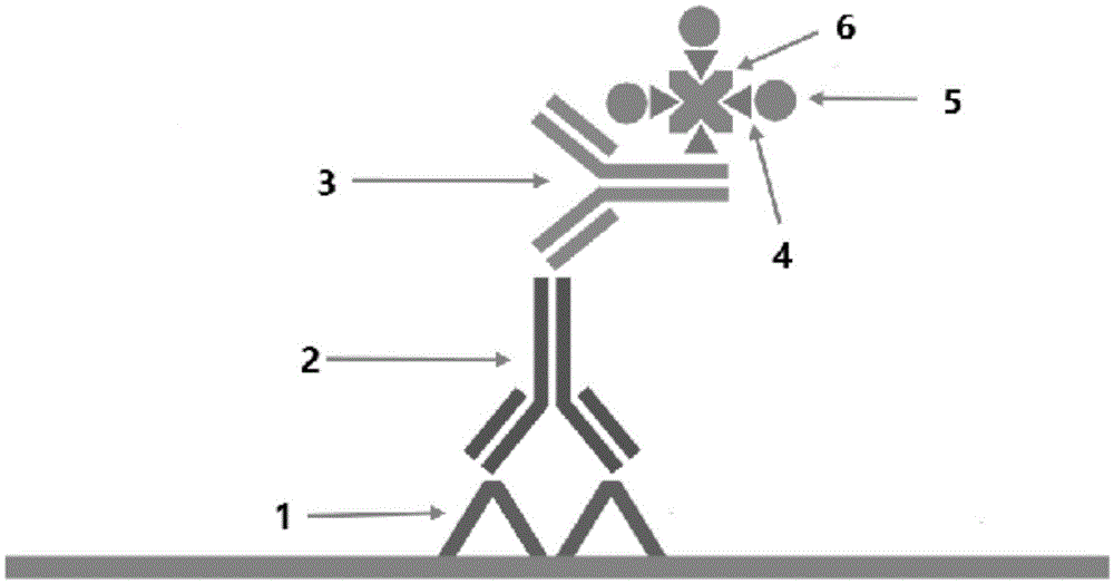
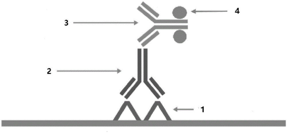
However, this method has a serious disadvantage: biotin is one of the vitamins that exist in the body, especially in the small intestine tissue, kidney tissue, brain tissue and liver tissue. Using the biotin-biophilin amplification method in the above There will be positive signals in the tissue that have nothing to do with the test itself, that is, the possibility of false positives and misdiagnosis
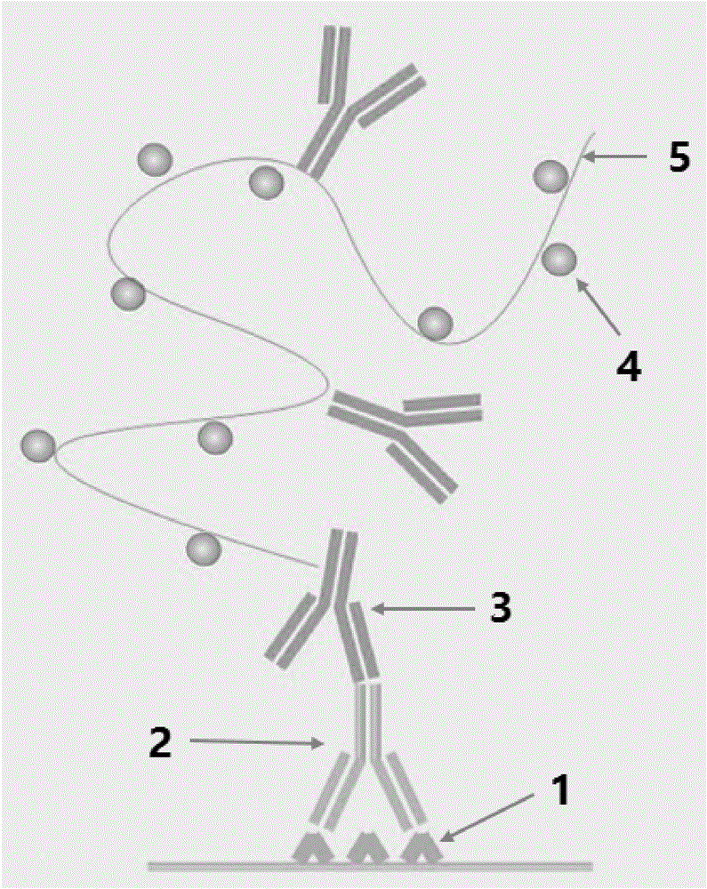
However, the disadvantages of this method are: the chain polymer enzyme-linked secondary antibody has a loose overall structure and a long volume, which affects the penetration of tissues, and it takes a long time to detect some characteristic substances existing in the nucleus. In order to achieve the required sensitivity, the signal amplification effect of some tissues fixed (soaked) in formalin for a long time is not effective enough

Method used


Image

  • Polymer enzyme-antibody and preparation method thereof
  • Polymer enzyme-antibody and preparation method thereof
  • Polymer enzyme-antibody and preparation method thereof

Examples


Embodiment 1

[0037] 1) Dissolve 2,000,000U peroxidase (SigmaChemicalCompany, Catalog#No.P8375-250KU, 7.5g) in 2.0ml of sodium bicarbonate buffer (0.1M, pH8.00) at room temperature, add 0.24mL of DVS (Sigma Chemical Company, Catalog#No.V3700-5G) then gently stirred at 30°C for 30 minutes;

[0038] 2) Purify the above reaction solution through a 50 ml SephadexG-25 column (Sigma Chemical Company, Catalog No. G25150) to remove the remaining DVS, and perform the purification in sodium bicarbonate buffer (0.1M sodium bicarbonate, pH 8.0). Collect the brown fraction, which is the activating enzyme;

[0039] 3) Concentrate the above activating enzyme to 4.0ml with a centrifugal concentration tube;

[0040] 4) Add 144 mg of PAMAMDendrimer 5.0 (Sigma Chemical Company, Catalog No. 536709-5G) to the above-mentioned concentrated 4 ml of activating enzyme (the molecular ratio of activating enzyme to PANMAM is 40:1). The solution was stirred gently at 30°C for 16 hours;

[0041] 5) Purify the reaction...

Embodiment 2

[0049] 1) Dissolve 2,000,000U peroxidase (SigmaChemicalCompany, CatalogNo.P8375-250KU, 7.5g) in 2.0ml of sodium bicarbonate buffer (0.1M, pH8.00) at room temperature, add 0.24mL of DVS (SigmaChemicalCompany , CatalogNo.V3700-5G) then stirred gently at 30°C for 30 minutes;

[0050] 2) Purify the above reaction solution through a 50 ml SephadexG-25 column (Sigma Chemical Company, Catalog No. G25150) to remove the remaining DVS, and perform the purification in sodium bicarbonate buffer (0.1M sodium bicarbonate, pH 8.0). Collect the brown fraction, which is the activating enzyme;

[0051] 3) Concentrate the above activating enzyme to 4.0ml with a centrifugal concentration tube;

[0052] 4) Add 144 mg of PAMAMDendrimer 5.0 (Sigma Chemical Company, Catalog No. 536709-5G) to the above-mentioned concentrated 4ml of activating enzyme (the molecular ratio of activating enzyme to PANMAM is 40:1), and put the solution in Stir gently for 16 hours at 30°C;

[0053] 5) Purify the reaction...


PUM


Abstract

The invention belongs to the technical field of medical detection, and especially relates to a polymer enzyme linked antibody and a preparation method thereof. The polymer enzyme is prepared by carrying out polymerization between DVS activated enzyme and PAMAN Dendrimer series branched organic molecular carriers. The provided method overcomes the main shortages of a conventional polymer enzyme linking method that takes chain-like polymer (glucan or polypeptide) as the carriers; moreover, the volume of polymer enzyme is greatly reduced; the density of enzyme labeling on antibody is increased, and the number of enzymes that is linked to the secondary antibody in a unit volume is largely increased. The produced polymer enzyme linked secondary antibody has the higher sensitivity and stability, compared with those of conventional enzyme labeled secondary antibody.

Description

technical field [0001] The invention belongs to the technical field of medical detection, and in particular relates to a polymer enzyme-antibody and a preparation method thereof. Background technique [0002] Immunohistochemistry is an immunohistochemical technique for detecting tumor-specific proteins or antigens in pathological specimens by using the specific recognition characteristics of antigens and antibodies[1,2]. This technology can not only detect the nature and quantity of the characteristic substance, but also because this technology can detect the original position of the characteristic protein or antigen and detect it on the basis of maintaining the original shape of the tissue, making it a good tool for pathological etiology analysis (especially Analysis of tumor pathology) is the most effective method, and has become one of the most commonly used clinical methods for tumor diagnosis, identification, differentiation and efficacy prediction. [0003] The experi...

Claims


Application Information

Patent Timeline
IPC IPC(8): C07K19/00C07K1/13C07K1/16C12N11/08G01N33/535G01N33/574G01N33/68
CPCC07K1/13C07K1/16C07K16/4241C07K2317/54C07K2317/55C07K2319/61C12N9/0065C12N9/16C12N9/80C12N11/08C12Y301/03001C12Y305/01005G01N33/535G01N33/574G01N33/6854
Inventor 郭金灿
Owner XIAMEN TALENT BIOMEDICAL TECH CO LTD
Learn More