Visual puncture biopsy treatment needle

A needle biopsy and needle technology, which is applied in the field of visual needle biopsy treatment needles, can solve the problems of inability to make a diagnosis, inability to obtain enough tissue samples, mistakes, etc.

Pending Publication Date: 2016-06-01
LANZHOU UNIVERSITY
View PDF10 Cites 11 Cited by
  • Summary
  • Abstract
  • Description
  • Claims
  • Application Information

AI Technical Summary

Problems solved by technology

These methods are relatively blind, time-consuming, or require the assistance of other equipment, and mistakes occur from time to time
Sometimes, due to bleeding during the puncture, the diagnosis and treatment must be terminated, or even converted to open surgery for emergency treatment; or the tissue lesion cannot be observed, sufficient tissue samples cannot be obtained, and the diagnosis cannot be made
[0003] In addition, micro-plastic surgery technology has developed rapidly in recent years, and soft tissue filling such as collagen, hyaluronic acid, and granular fat has become the most cosmetic treatment. However, when injecting filling or extracting granular fat, the current blindness is not only easy to puncture blood vessels , cause bleeding, and it is easy to inject the above-mentioned filling materials through the vascular break, resulting in vascular embolism, blindness, hemiplegia and other serious consequences
[0004] In order to solve these problems, the visual puncture needle device (CN201668471U) uses a puncture needle with a camera and a liquid crystal display at the front end to realize visual puncture, but because there is no operation channel in the needle core, it cannot be performed under direct vision Operations such as obtaining tissue samples or hemostasis
A visible puncture needle and its visible puncture tube insertion needle (CN201160881Y) are provided with a tube insertion cavity, which can be used as a needle or injection after tube placement, but the tube cavity is too small to be inserted into an instrument for operation, and the cold light source used Both the image and the image are transmitted through optical fiber, and the image is connected and displayed in the rear end through the image receiving sensor and the image magnification display, but the image is a non-digital signal, which cannot be stored and transmitted
The visual puncture needle for thoracoabdominal surgery (CN203138637U) has 6-9 lumens in the needle body, which are used to insert the guide wire or laser beam to avoid entanglement, and can determine which lumen can be used to better diagnose and treat lesions. But its design must use a thicker needle body, which is not conducive to minimally invasive operation and lacks practicability
All above-mentioned inventions also have the shortcoming that can not stop bleeding at any time

Method used


Image

  • Visual puncture biopsy treatment needle
  • Visual puncture biopsy treatment needle
  • Visual puncture biopsy treatment needle

Examples


Embodiment 1

[0027] Embodiment 1 Visual puncture biopsy treatment needle assembly

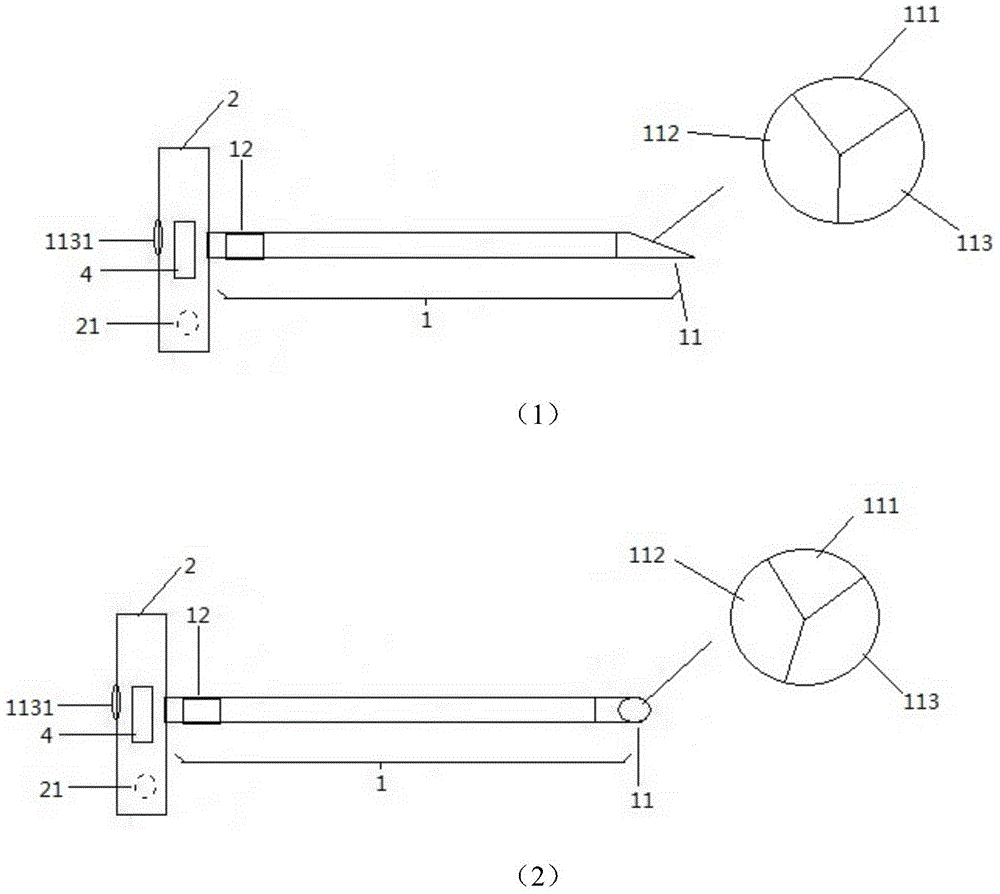
[0028] The present invention provides a visual puncture biopsy treatment needle, which comprises a needle body 1, a digital control terminal 2 and a display 3, wherein the digital control terminal 2 is connected to the needle body 1, and the display 3 is connected to the digital control terminal 2; the needle body 1 It is made of metal, and the outer surface of the middle part is coated with an insulating layer. The 0.5-5.0mm part of the front end of the needle body and the rear end of the needle body has no insulation layer.

[0029] The needle body 1 is divided into a data line tunnel 111, a wire tunnel 112 and 1-2 operation tunnels 113, and the three are arranged in a triangle, a quadrangle, or a way that the wire tunnel 112 is located in the middle. The built-in data line of data line tunnel 111, data line connects the miniature camera that needle body front end 11 places are provided with;

[0030] Th...

Embodiment 2

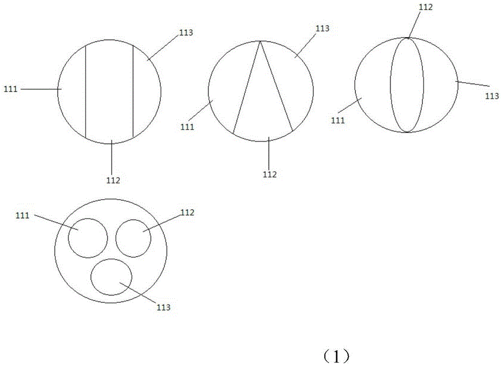
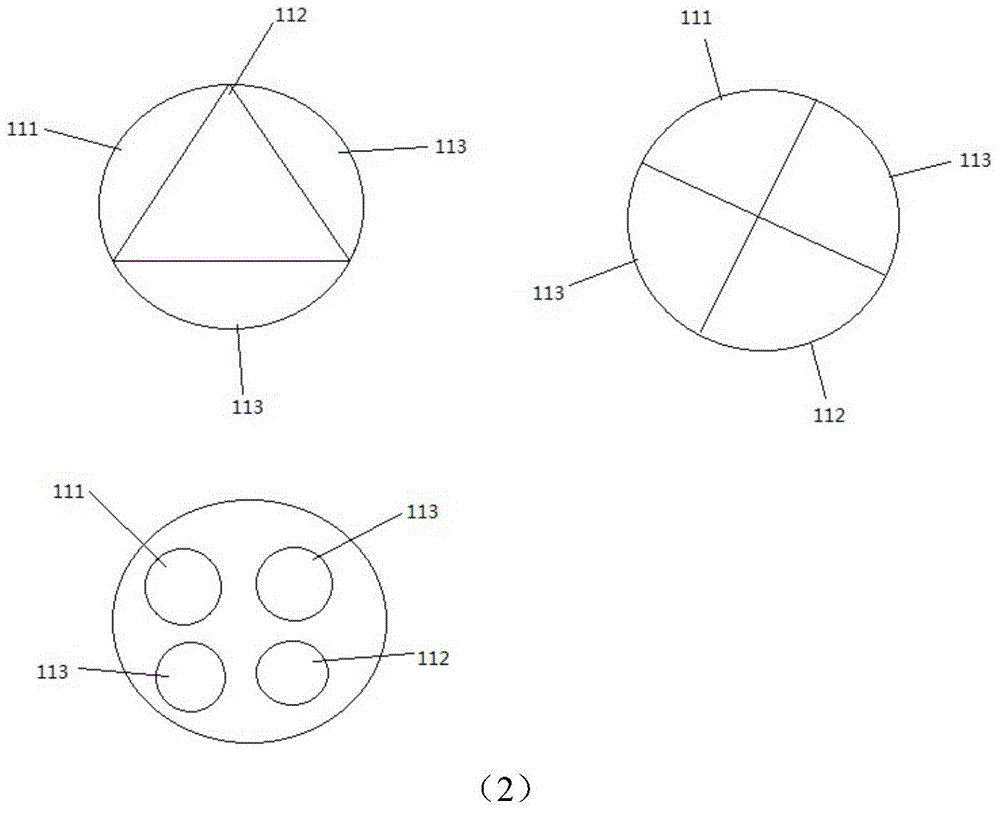
[0034] The distribution of each aperture in the needle body in embodiment 2

[0035] In the needle body 1 of the visible puncture biopsy treatment needle of the present invention, the data line hole 111, the wire hole 112 and the operation hole 113 can be divided and distributed according to actual needs, and the preferred technical solution is that the wire hole 112 is located in the middle of the needle body 1 , in order to better provide lighting for surgery.

[0036] (1) figure 2 (1): 1 data hole, 1 wire hole, 1 operation hole

[0037] (2) figure 2 (2): 1 data hole, 1 wire hole, 2 operation holes

Embodiment 3

[0038] Embodiment 3 visual puncture biopsy treatment needle is used for hemostasis or cutting

[0039] When using the present invention, an epidural puncture needle core or a scalp needle core for children or a heart floating catheter needle core or an interventional catheter needle core, etc. can be inserted in the operation hole (when the needle is blunt, first puncture the skin with a common sharp needle, Then use the present invention), and then puncture. During the puncture process, water can be injected through the operation hole for flushing and suction at any time to clear the field of vision, and at the same time, the metal part at the back end of the needle body can be electrocoagulated to stop bleeding or cut; it can also be rinsed through one operation hole, and electrodes or laser optical fibers can be placed in the other operation hole. After observing subcutaneous hemangioma and other diseased tissues, electrocoagulation or cutting or laser coagulation and gasif...


PUM


Abstract

The invention relates to the field of puncture diagnosis and treatment operation equipment, in particular to a visual puncture biopsy treatment needle. The treatment needle comprises a needle body 1, a digital control end 2 and a display 3, wherein the needle body 1 is made of metal, the outer surface of the middle is coated with an insulation layer, and the front end 11 and the back end 12 of the needle body are not coated with insulation layers. The treatment needle can be used for electrotome cutting or electric coagulation hemostasis while performing visual puncture diagnosis and treatment operation, integrates electrotome, endoscope, biopsy and treatment and is simple in structure, diversified in function and convenient to use.

Description

technical field [0001] The invention relates to the field of puncture diagnosis and treatment operation equipment, in particular to a visual puncture biopsy treatment needle. Background technique [0002] At present, minimally invasive technology strives to complete the diagnosis and treatment of lesions in the body with the smallest incision path and the least tissue damage. It has the characteristics of less bleeding, less postoperative pain, faster recovery, and minimal or no scars. Clinically, puncture and biopsy of subcutaneous tissue, kidney, liver, etc., or cone drainage, or puncture injection of drugs, or small needle-knife treatment (cervical spondylosis, lateral epicondylitis of humerus, stenosing tenosynovitis of flexor tendon, Three lumbar transverse process syndrome, chronic lumbar muscle strain, etc.), or foreign bodies (broken needles, broken catheters and other foreign bodies) are taken out, it may be necessary to rely on hand feeling, or predict the depth u...

Claims


Application Information

Patent Timeline
Patent Type & Authority Applications(China)
IPC IPC(8): A61B10/02A61B18/12A61B18/22A61M1/00A61M5/32
Inventor 张选奋余祎蒙礼娟乔建雄
Owner LANZHOU UNIVERSITY
Why Eureka
Try Eureka