Hydrogel microsphere fluorescence sensor as well as preparation method and application thereof

A fluorescence sensor, hydrogel microsphere technology, applied in fluorescence/phosphorescence, instruments, scientific instruments, etc., can solve the problems of rapid detection of limited ions, harsh detection requirements, expensive detection costs, etc., to achieve high sensitivity and great application prospects , the effect of simple steps

Inactive Publication Date: 2016-07-20
NANJING UNIV OF TECH
View PDF7 Cites 15 Cited by
  • Summary
  • Abstract
  • Description
  • Claims
  • Application Information

AI Technical Summary

Problems solved by technology

However, the rapid detection of ions is limited due to expensive det

Method used


Image

  • Hydrogel microsphere fluorescence sensor as well as preparation method and application thereof
  • Hydrogel microsphere fluorescence sensor as well as preparation method and application thereof
  • Hydrogel microsphere fluorescence sensor as well as preparation method and application thereof

Examples


Embodiment 1

[0045] Embodiment 1 Preparation of hydrogel precursor solution and oil phase and preparation of microspheres

[0046] 3g PEG-DA, 2.7g HEPES buffer and 0.3g photoinitiator (2-hydroxy-2-methyl-1-phenyl-1-propanone) were thoroughly mixed to prepare a hydrogel solution. The fluorescent probe was dissolved in DMSO to a concentration of 1mMol / L. The probe solution was diluted and mixed with the DMSO solution of the probe and the HEPES buffer according to the volume ratio of 1:5. The hydrogel precursor solution was fully mixed with the hydrogel solution and the probe solution at a volume ratio of 1:5, and was ultrasonically prepared. The oil phase consists of 60%-99% organic solvents including n-decane, n-hexane, n-octane or n-butane, etc., mixed with 10-25% oil-soluble surfactants such as Span20, Span40, Span60, Span80, Tween85 or Alkyl tertiary amine salts, etc., mixed thoroughly, ultrasonically removed air bubbles, set aside.

[0047]Use two syringes to absorb the appropriate a...

Embodiment 2

[0048] The microsphere sensor that embodiment 2 makes is to the selectivity of metal ion

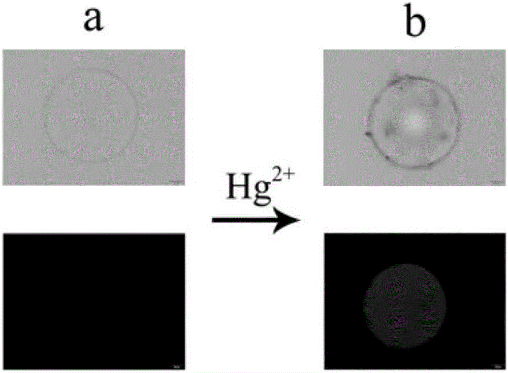
[0049] Take the microsphere sensor prepared in Example 1, draw one and place it in a 96-well plate, then add mercury ions, let it stand at room temperature for 5 minutes, and take optical images and fluorescence images under a fluorescence microscope. The result is as figure 1 shown.

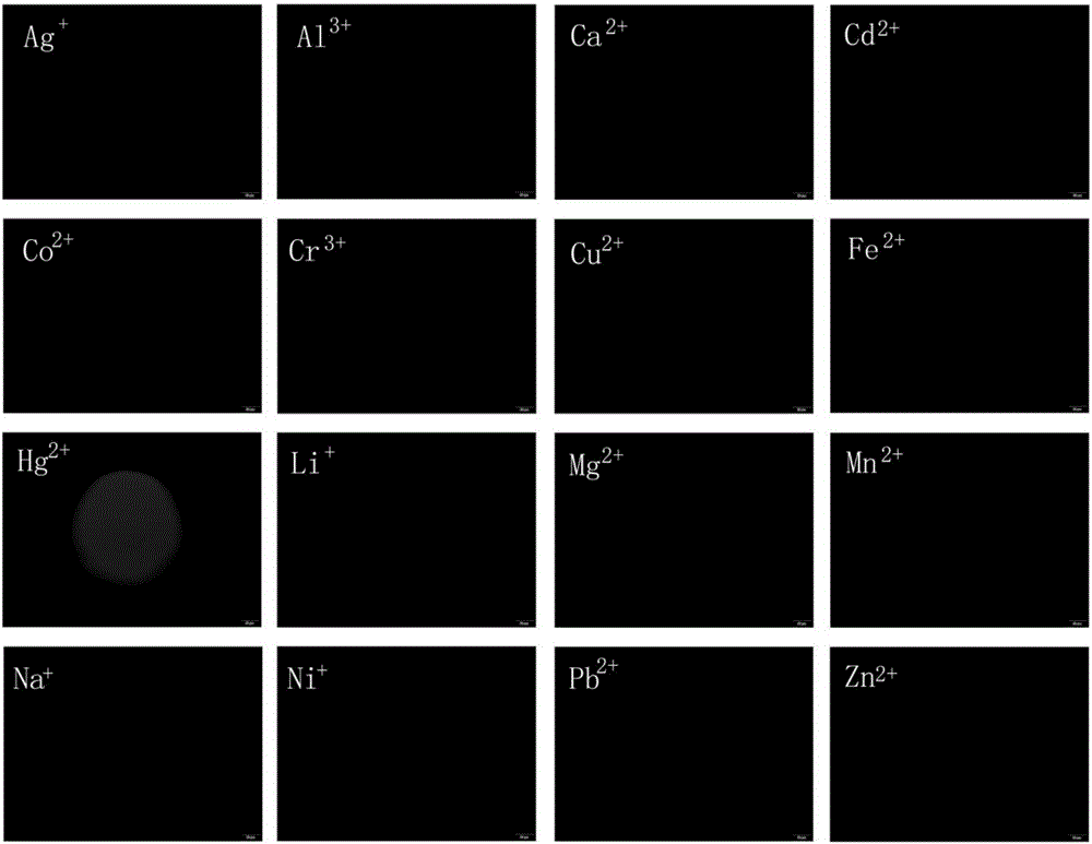
[0050] Prepare the following metal ion solutions: Cu 2+ ,Pb 2+ , Na + ,Hg 2+ , Fe 2+ ,Mn 2+ ,Cr 3+ ,Co 2+ ,Zn 2+ ,Mg 2+ , Li + , Ni 2+ ,Cd 2+ , Ag + , Ca 2+ and Al 3+ , the concentration is 1 μMol / L. Select 16 wells on a 96-well plate and inject 0.2 mL each of the above metal ions. Then, put one microsphere of Example 1 into each well, let it stand for 5 minutes, and take a fluorescent image under a fluorescent microscope, and the conditions such as exposure time and light intensity are uniform. The result is as figure 2 shown.

[0051] The above results show that: the microsphere sen...

Embodiment 3

[0052] Fluorescence analysis of the microsphere sensor and mercury ion made in embodiment 3

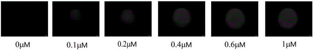
[0053] Prepare mercury ions at 0 μM, 0.1 μM, 0.2 μM, 0.4 μM, 0.6 μM, and 1.0 μM. Pipette 0.2 mL of the above-mentioned mercury ion solution into a 96-well plate, and then put microsphere sensors in each well, let it stand for 5 minutes, and take fluorescence images under a fluorescence microscope. The result is as image 3 shown.

[0054] The relationship between mercury ion concentration and fluorescence intensity. The fluorescence microscope selects the grayscale mode, and then takes grayscale pictures at mercury ion concentrations of 0μM, 0.2μM, 0.4μM, 0.6μM and 0.8μM, and calculates the grayscale value in the same area of ​​each picture, which is the fluorescence intensity. Make a graph of concentration versus fluorescence intensity. The result is as Figure 4 shown.

[0055] The microsphere sensor of Example 1 was placed in the wells of the 96-well plate, placed on the obse...


PUM

PropertyMeasurementUnit
Diameteraaaaaaaaaa
Diameteraaaaaaaaaa

Abstract

The invention discloses a hydrogel microsphere fluorescence sensor as well as a preparation method and application thereof. By use of the advantages of a hydrogel material, the hydrogel material is applied to wrapping a fluorescence probe; the hydrogel is fully mixed with the fluorescence probe, and the mixture is directly processed into a fluorescence sensor system which has a microsphere structure and is applicable to fluorometric analysis and detection by virtue of an ultraviolet polymerization reaction; successfully cured hydrogel micropheres have favorable flexible structures and can not release hydrogel monomers without influencing detection; due to a net-shaped structure, the wrapped fluorescence probe is quite stably dispersed in the hydrogel with out leaking through the micropheres; detection substances can freely enter into or move out of the whole microsphere fluorescence sensor system; in combination with a fluorescence microscope, the hydrogel microsphere fluorescence sensor is capable of quite conveniently analyzing and detecting ions, molecules and other relevant substances; the hydrogel microsphere fluorescence sensor is a microsphere which is formed by wrapping the fluorescence probe with hydrogel and has a diameter of 300-500 mu m.

Description

technical field [0001] The invention relates to a sensor, a preparation method and an application, more specifically to a hydrogel microsphere fluorescent sensor, a preparation method and an application. Background technique [0002] In conventional fluorescent sensor systems, fluorescent probes are suspended in aqueous solutions or immobilized on solid substrates. When the detection substance is added, the fluorescent probe dispersed in the water phase can detect whether there is a target molecule. If there is a target molecule, the fluorescent probe will show a very direct fluorescence change. However, there are certain limitations. For example, the fluorescent probes are relatively scattered in the entire detection system, and the detection sensitivity is average; sometimes the color of the detection object is more complex, and once the fluorescent probe is added to the detection object, it cannot be separated, and it is difficult to carry out Fluorescence analysis dete...

Claims


Application Information

Patent Timeline
IPC IPC(8): G01N21/64
CPCG01N21/6428G01N2021/6497
Inventor 陈小强宋振虎王芳魏廷文强健
Owner NANJING UNIV OF TECH
Why Eureka
Try Eureka