Preparation method of single chain antibody-modified targeting micro-bubble ultrasonic contrast agent

A technology of ultrasound contrast agent and single-chain antibody, which is applied in the fields of ultrasound molecular imaging and biomedical engineering, can solve the problems of low output efficiency of targeted ultrasound contrast agents, and achieve target specificity, high link rate, and response The effect of simple process and conditions

Active Publication Date: 2016-08-17
THE THIRD AFFILIATED HOSPITAL OF SUN YAT SEN UNIV
View PDF7 Cites 1 Cited by
  • Summary
  • Abstract
  • Description
  • Claims
  • Application Information

AI Technical Summary

Problems solved by technology

[0010] The purpose of the present invention is to address the defect of low production efficiency of existing targeted ultrasound contrast agents, and propose an improved method based on the thiol-maleimide method and linking antibodies to prepare targeted micro-contrast agents with high yields. bubble ultrasound contrast agent

Method used


Image

  • Preparation method of single chain antibody-modified targeting micro-bubble ultrasonic contrast agent
  • Preparation method of single chain antibody-modified targeting micro-bubble ultrasonic contrast agent
  • Preparation method of single chain antibody-modified targeting micro-bubble ultrasonic contrast agent

Examples


Embodiment 1

[0064] Example 1: Preparation of targeted microbubble ultrasound contrast agent by a new method

[0065] Weigh the phospholipid powder DPPC, DPPA, DSPE-PEG according to the mass fraction ratio of 7:1:2 2000 -A total of 20 mg of maleimide was placed in a small beaker, and 4 mL of chloroform was added. Heat to 50°C in a water bath, stir with a glass rod until the phospholipid is completely dissolved; then transfer the phospholipid chloroform solution to a round bottom flask with a volume of 50 mL. The chloroform was completely evaporated on a rotary evaporator (the temperature of the water bath was 50° C. and the rotation speed was 120 rpm), and a phospholipid film was formed at the bottom of the round-bottomed flask. Add hydration solution (PBS) into the round bottom flask, seal it with plastic wrap and rubber band, and hydrate in a constant temperature shaker at 60° C. and 120 rpm for 45 minutes to obtain a liposome solution. Maleimidated liposomes (mal-liposome) can be obta...

Embodiment 2

[0073] Example 2: Efficacy detection of microvesicle-linked single-chain antibody

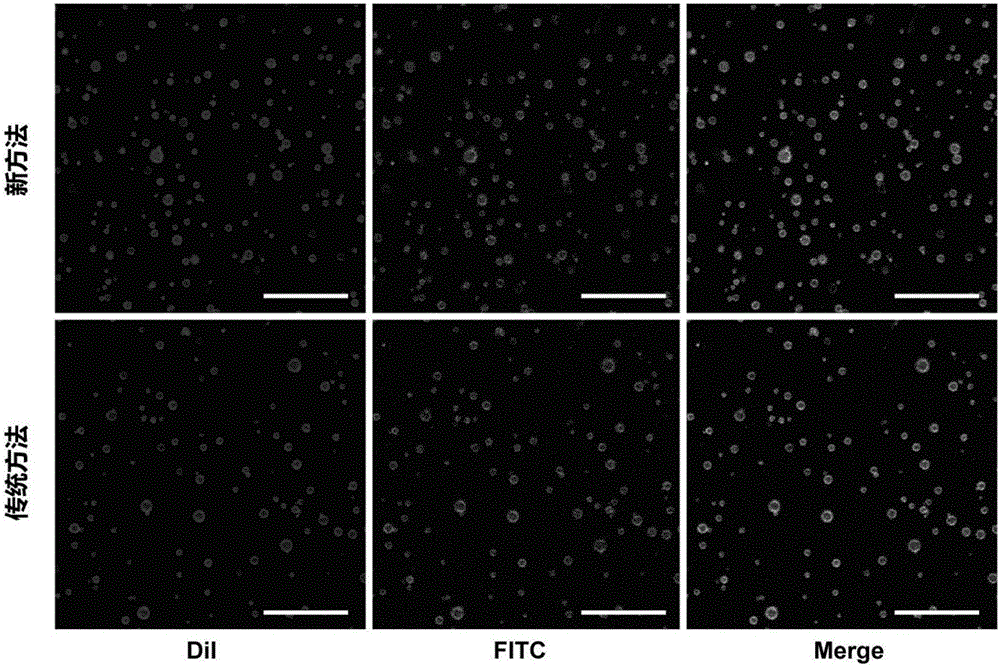
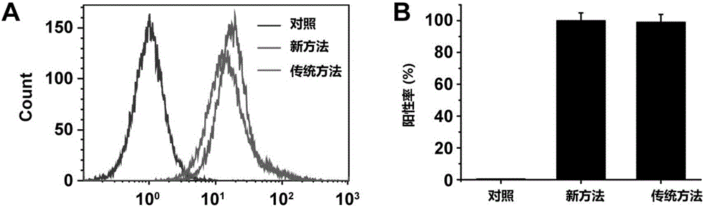
[0074] In this example, the preparation of MB by the "new method" was observed qualitatively and quantitatively through laser confocal microscopy and quantitative flow cytometry. ICAM-1 The basic potency and the preparation of MB with "traditional method" ICAM-1 comparative advantage. The specific method is as follows: in the preparation of MB ICAM-1 When forming a lipid film with phospholipids, add 0.5mg of hydrophobic red fluorescent dye DiI for labeling MB ultrasonic contrast agent; ICAM-1 After that, the MB ICAM-1 Incubate with the green fluorescent dye FITC-labeled donkey anti-rabbit fluorescent secondary antibody, centrifuge and wash to obtain MB labeled with red fluorescence and scICAM-1 labeled with green fluorescence ICAM-1 .

[0075] Using laser confocal microscopy to observe the preparation of MB by two methods ICAM-1 Red fluorescently labeled MB surface linked to green fluores...

Embodiment 3

[0076] Example 3: Detection of targeted microbubble ultrasound contrast agent concentration and ultrasound imaging ability

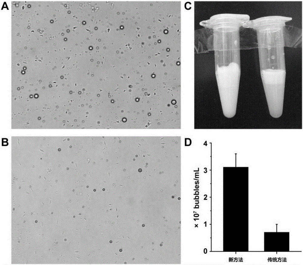
[0077] This embodiment prepares MB by detecting "new method" and "traditional method" ICAM-1 The final output rate highlights the advantages of the "new method" of the present invention for preparing targeted microbubble ultrasound contrast agents. The specific methods are as follows: ①Directly observe the two methods to prepare MB ICAM-1 of the final product. The results are attached image 3 As shown in A, MB prepared by two methods ICAM-1 All the EP tubes are milky white liquid that is layered. Since MB is a hollow microbubble structure, after standing still (about 15 minutes), MB floats up to cause solution layering (the upper layer with high MB concentration is milky white, and the lower layer with low MB concentration is translucent). Regardless of the milky white liquid on the upper layer or the translucent liquid on the lower layer, the MB pr...


PUM


Abstract

The invention belongs to the field of medical science, and relates to a single chain antibody-modified targeting micro-bubble ultrasonic contrast agent and a preparation method thereof. The preparation method comprises the following steps: preparing a maleimidized liposome; opening the disulfide bond of an antibody by using a reducing agent to form a mercapto group-exposed single chain antibody; linking the single chain antibody to the surface of the liposome to form a targeting liposome; and preparing the targeting micro-bubble ultrasonic contrast agent through a mechanical oscillation technology. The method can effectively improve the yield of the targeting micro-bubble ultrasonic contrast agent, and has an important application prospect in the invention and in relevant targeting micro-bubble ultrasonic contrast agent industrial production processes.

Description

technical field [0001] The invention relates to the fields of ultrasonic molecular imaging and biomedical engineering, in particular to a method for preparing a lipid microbubble ultrasonic contrast agent modified by a single-chain antibody. technical background [0002] Ultrasound molecular imaging is a molecular imaging method that uses targeted microbubble contrast agents as molecular probes and uses contrast-enhanced ultrasound as an imaging mode to conduct qualitative and quantitative research on the biochemical processes in vivo at the cellular and molecular levels. Accuracy and sensitivity have important application prospects in the fields of cardiovascular, inflammatory, tumor and other diseases. Among them, targeted ultrasonic molecular probes are the key to ultrasonic molecular imaging technology. Ultrasound molecular probes reported in the literature are mainly microbubble ultrasound contrast agents: specific ligands (antibodies, polypeptides, nucleic acid fragme...

Claims


Application Information

Patent Timeline
Patent Type & Authority Applications(China)
IPC IPC(8): A61K49/22
CPCA61K49/221A61K49/223
Inventor 尹庭辉郑荣琴邱晨游宇佳王平任杰
Owner THE THIRD AFFILIATED HOSPITAL OF SUN YAT SEN UNIV
Why Eureka
Try Eureka