Preparation method of marrow fluid smear
A technique for bone marrow fluid and bone marrow, applied in the field of preparation of bone marrow fluid smears, can solve the problems of uneven coloring of bone marrow smears, unfavorable bone marrow cell examination, too deep or too light staining, etc. Vivid, good contrast effect
- Summary
- Abstract
- Description
- Claims
- Application Information
AI Technical Summary
Problems solved by technology
Method used
Image
Examples
Embodiment 1
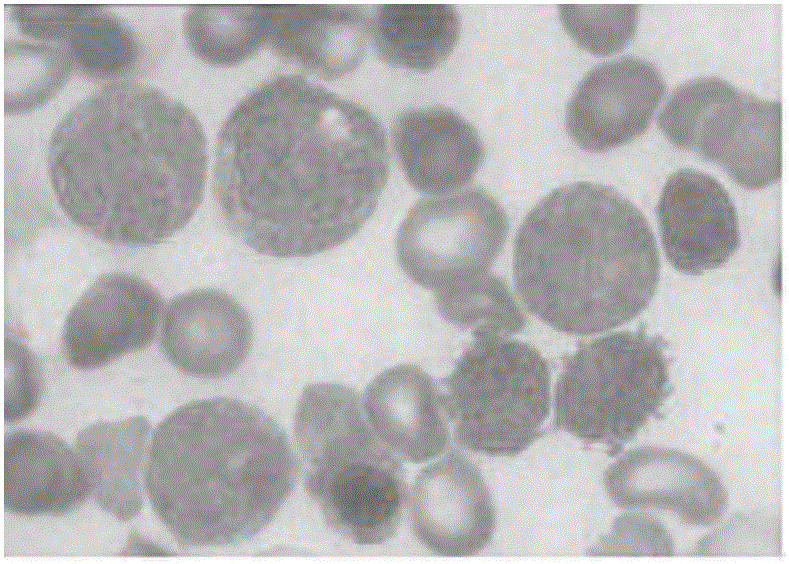
[0031] Embodiment 1, the preparation of a kind of bone marrow fluid smear
[0032] S1 Extract the bone marrow fluid, add 0.2% of the volume of the bone marrow fluid, and react for 35 seconds with an anticoagulant at a concentration of 2 mg / ml. The anticoagulant consists of heparin and dipotassium edetate in a weight ratio of 1:2. Obtain anticoagulated bone marrow fluid;
[0033] S2 Take 5 μL of the anticoagulated bone marrow liquid obtained in step S1 and drop it onto one end of the glass slide, and smear the bone marrow liquid into a thin slice with a push slide, the angle between the push slide and the slide glass is 35°, and a smear is obtained;
[0034] S3 arranges the smears obtained in step S2 on the slide rack, puts the slide rack into the staining vat 1 and fixes it for 1 min, and the staining vat 1 is a staining liquid, and the staining liquid is 1 g of Wright's dye, Add 0.3g of Giemsa dye powder to 500ml of methanol, then add 10ml of glycerin and mix well, place the...
Embodiment 2
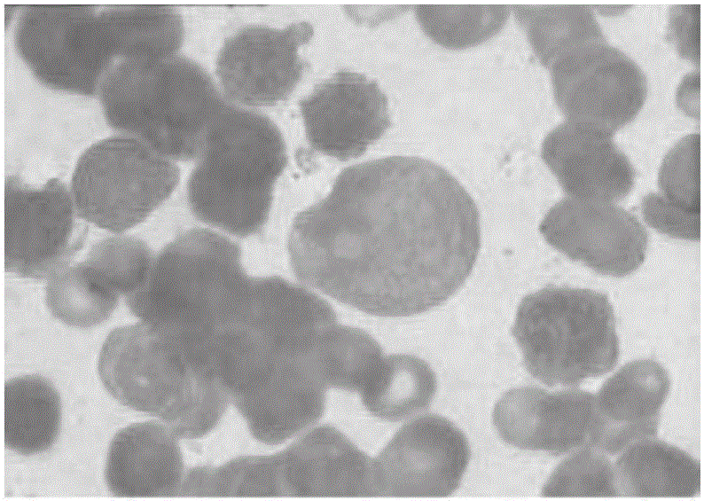
[0037] Embodiment 2, the preparation of a kind of bone marrow fluid smear
[0038] S1 Extract the bone marrow fluid, add 0.2% of the volume of the bone marrow fluid, and react for 45 seconds with an anticoagulant with a concentration of 3 mg / ml. The anticoagulant consists of heparin and dipotassium edetate in a weight ratio of 1:3. Obtain anticoagulated bone marrow fluid;
[0039] S2 Take 6 μL of the anticoagulated bone marrow liquid obtained in step S1 and drop it on one end of the glass slide, and use a push slide to smear the bone marrow liquid into a thin slice, and the angle between the push slide and the slide glass is 35° to obtain a smear;
[0040] S3 arranges the smears obtained in step S2 on the slide rack, puts the slide rack into the staining vat 1 and fixes it for 2 min, and the staining vat 1 is a staining liquid, and the staining liquid is 1 g of Wright's dye, Add 0.3g of Giemsa dye powder to 500ml of methanol, then add 10ml of glycerin and mix well, place the ...
Embodiment 3
[0043] Embodiment 3, the preparation of a kind of bone marrow fluid smear
[0044] S1 Extract the bone marrow fluid, add 0.3% of the volume of the bone marrow fluid, and react for 55 seconds with an anticoagulant at a concentration of 4 mg / ml. The anticoagulant consists of heparin and dipotassium edetate in a weight ratio of 1:4. Obtain anticoagulated bone marrow fluid;
[0045] S2 Take 7 μL of the anticoagulated bone marrow liquid obtained in step S1 and drop it on one end of the glass slide, and use a push slide to smear the bone marrow liquid into a thin slice, and the angle between the push slide and the slide glass is 35° to obtain a smear;
[0046] S3 arranges the smears obtained in step S2 on the slide rack, puts the slide rack into staining vat 1 and fixes it for 3 min, and the staining vat 1 is a staining liquid, and the staining liquid is 1 g of Wright's dye, Add 0.3g of Giemsa dye powder to 500ml of methanol, then add 10ml of glycerin to mix well, place the prepare...
PUM
 Abstract
Description
Claims
Application Information
 - R&D
- Intellectual Property
- Life Sciences
- Materials
- Tech Scout
- Unparalleled Data Quality
- Higher Quality Content
- 60% Fewer Hallucinations
Browse by: Latest US Patents, China's latest patents, Technical Efficacy Thesaurus, Application Domain, Technology Topic, Popular Technical Reports.
© 2025 PatSnap. All rights reserved.Legal|Privacy policy|Modern Slavery Act Transparency Statement|Sitemap|About US| Contact US: help@patsnap.com


