Method of enriching and detecting miRNA in sample on the basis of magnetic bead coupled antibody, and matched microfluidic apparatus

A microfluidic device, a technology in samples, applied in chemical instruments and methods, biochemical equipment and methods, laboratory containers, etc., to achieve high-efficiency reaction and enrichment effects

Inactive Publication Date: 2017-03-15
EAST CHINA UNIV OF SCI & TECH
View PDF2 Cites 3 Cited by
  • Summary
  • Abstract
  • Description
  • Claims
  • Application Information

AI Technical Summary

Problems solved by technology

The four Argonaute proteins can recognize different small RNA molecules and form the RNA-induced silencing complex (RISC), but so far only the Ago2-miRNA complex has been shown to have cleavage activity for target RNA cleavage

Method used


Image

  • Method of enriching and detecting miRNA in sample on the basis of magnetic bead coupled antibody, and matched microfluidic apparatus
  • Method of enriching and detecting miRNA in sample on the basis of magnetic bead coupled antibody, and matched microfluidic apparatus
  • Method of enriching and detecting miRNA in sample on the basis of magnetic bead coupled antibody, and matched microfluidic apparatus

Examples


Embodiment 1

[0048] Example 1——enrichment of Ago2-miRNA complex in serum and detection of miRNA

[0049] 1 Preparation of immunomagnetic beads:

[0050] 1.1 Take 1.5 mg of Protein G magnetic beads and place them in a 2 mL centrifuge tube, use a magnetic separator to remove the original storage solution, and use 200 μL of PBS buffer to wash twice, and use a magnetic separator to remove the cleaning solution;

[0051] 1.2 Use 200 μL of PBS buffer to resuspend the magnetic beads, add 8 μg of anti-Ago2 rabbit monoclonal antibody, and incubate on an incubator at room temperature for 15 min;

[0052] 1.3 Wash the magnetic beads twice with 200 μL PBST buffer, shake gently for 1 min each time to prevent the beads from coagulating, remove excess antibody at the same time and transfer to a new centrifuge tube to obtain anti-Ago2 immunomagnetic beads.

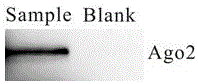
[0053] 2. Immunomagnetic beads enrich the Ago2-miRNA complex in serum and detect miRNA (magnetic beads without antibody coupling are used as negative ...

Embodiment 2

[0061] Example 2—Microfluidic device enriches Ago2-miRNA complex in serum and detects miRNA

[0062] 1 The preparation of immunomagnetic beads is the same as in Example 1.

[0063] 2 Microfluidic device preparation:

[0064] 2.1 Design and prepare the microfluidic chip template according to the above magnetic bead immunoenrichment method;

[0065] 2.2 Pouring PDMS polymer on the microfluidic chip template and curing to obtain PDMS membrane;

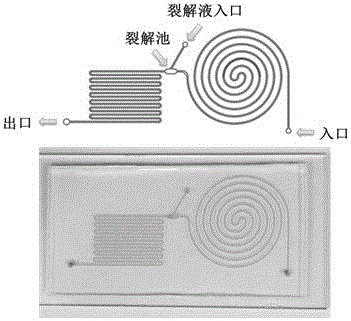
[0066] 2.3 Plasma bonding the PDMS membrane and the glass sheet, and packaging to obtain the required microfluidic chip device ( image 3 , Figure 4 ).

[0067] 3 Immunomagnetic beads were used to enrich Ago2-miRNA complexes in serum and detect miRNA on the microfluidic platform (the experiment was performed without antibody-coupled magnetic beads as a negative control)

[0068] 3.1 Pass 1 mL of BSA solution with a concentration of 1% through the microfluidic channel, the flow rate is controlled at 50 μL / min, and the microfluidic ch...


PUM


Abstract

The invention discloses a method of enriching and detecting miRNA in a sample on the basis of a magnetic bead coupled antibody, and a matched microfluidic apparatus. The method herein includes the following steps: 1) connecting an anti-Ago2 monoclonal antibody to magnetic beads to obtain anti-Ago2 immunomagnetic beads; 2) mixing the anti-Ago2 immunomagnetic beads with a sample to enrich an Ago2-miRNA composite through an antigen-antibody immuno-reaction; 3) by means of a lysis liquid, carrying out high-temperature transformation to the enriched Ago2-miRNA composite so as to release miRNA for subsequent quantitative analysis. In the method, by means of combination of high-specificity of antigen-antibody immunological recognition and combination, simplicity and operability of high-affinity magnetic bead separation and high-effective reaction and enrichment properties of the microfluidic apparatus, various miRNAs, which are related to diseases, especially tumors, in bio-samples, such as blood, urine, saliva, synovial fluid, cell lysis liquid and the like, can be quickly detected. The method has important significance on early and accurate diagnosis of clinically severe diseases.

Description

technical field [0001] The invention relates to a method for enriching and detecting miRNA in a biological sample and a supporting microfluidic device, belonging to the technical field of high-sensitivity detection of micro or trace microRNA in samples and early rapid diagnosis of major diseases such as tumors. Background technique [0002] MicroRNA (miRNA) is a class of non-coding RNA involved in the regulation of post-transcriptional gene expression in vivo, and plays an important role in various physiological and pathological processes. There are significant differences in miRNA expression profiles between tumor cells and normal tissue-derived cells. About 50% of the annotated miRNA genes are located at vulnerable gene loci related to tumor formation, which shows that miRNAs play a very important role in the occurrence of tumors. effect. Therefore, it is of great significance to study the types and contents of miRNAs for the diagnosis, classification and treatment of tum...

Claims


Application Information

Patent Timeline
Patent Type & Authority Applications(China)
IPC IPC(8): C12Q1/68B01L3/00
CPCC12Q1/6806B01L3/5027C12Q1/6851C12Q1/6886C12Q2600/178C12Q2527/125C12Q2563/143C12Q2563/149C12Q2531/113C12Q2563/159C12Q2565/629
Inventor 叶邦策吴姗左鹏
Owner EAST CHINA UNIV OF SCI & TECH
Why Eureka
Try Eureka