
Novel degradable supporter for intestinal canal anastomosis surgery

A support and surgical technology, applied in the field of surgical tools, can solve the problems of leakage of intestinal contents, cumbersome operation, and difficult suturing, so as to shorten the operation time, increase the suturing speed, and reduce the damage of the intestinal wall

Inactive Publication Date: 2017-04-19
NORTHEAST AGRICULTURAL UNIVERSITY
View PDF6 Cites 0 Cited by
  • Summary
  • Abstract
  • Description
  • Claims
  • Application Information

AI Technical Summary

Problems solved by technology

At present, there are many methods of digestive tract anastomosis, such as manual suture, stapler anastomosis, laser welding, adhesive anastomosis, etc.; traditional intestinal end-to-end anastomosis is mostly performed after partial resection of the necrotic intestinal tract, and aligning the healthy intestinal tract with surgical silk or catgut. Double-layer suture, cumbersome operation, easy to cause intestinal stenosis in puppies with thin intestinal tubes
Due to the empty intestinal cavity, the intestinal wall is close together, suturing is difficult, and the anastomotic effect is not good. After the operation, it is easy to cause leakage of intestinal contents or intestinal stenosis, and even lead to the failure of the operation
And if the suturing time is too long, the serosa layer of the intestinal wall will suffer more mechanical stimulation, and adhesions will easily occur, which is not conducive to the recovery of digestive function after surgery.
[0003] In the medical field, degradable one-piece intestinal anastomosis aids and intestinal anastomosis stents have been developed, but these devices are too large in diameter for small animals and expensive, and are not suitable for small animal intestinal anastomosis operations

Method used


Image

  • Novel degradable supporter for intestinal canal anastomosis surgery
  • Novel degradable supporter for intestinal canal anastomosis surgery
  • Novel degradable supporter for intestinal canal anastomosis surgery

Examples


Embodiment 1

[0027] Example 1 Preparation of support

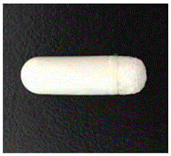
[0028] The starch puffed product is made hollow, with one end closed and one end open. Fill a group of puffs (generally 2) with 0.2 g of enrofloxacin, 0.25 g of amoxicillin and 0.01 g of diclofenac sodium. Heat the bone wax in a water bath (100°C) to melt it. Completely immerse a group of puffed products in the melted liquid bone wax. After the air in it is discharged, take out the puffed products and place them under the ultraviolet light for disinfection. After they cool naturally, repeat immersion in the liquid bone wax once, and then use ultraviolet light after cooling. The lamp is irradiated and sterilized, and put into a sterile sealed bag for use. The prepared supports are as follows: figure 1 .

Embodiment 2

[0029] Example 2 Preparation of support

[0030] The experimental method is the same as that of Example 1, the only difference being: filling enrofloxacin (0.2g), amoxicillin (0.2g) and diclofenac sodium (0.01g) into a group of puffs (generally 2).

Embodiment 3

[0031] The preparation of embodiment 3 supports

[0032] The experimental method is the same as that of Example 1, the only difference is that enrofloxacin (0.25g), amoxicillin (0.2g) and diclofenac sodium (0.02g) are filled into a group of puffs (generally 2).


PUM


Abstract

The invention provides a degradable supporter for intestinal canal anastomosis surgery, wherein hollow starch expanded material is used as the main body of the supporter and the expanded material is filled with antibiotic medicine and analgesic, the supporter is prepared by sealing the supporter main body with bone wax. A series of scientific component experiments is carried out through shaping the starch expanded material, integrating multiple medicaments and preparing the external protection layer so that the supporter is provided with features such as supporting, drug therapy and timing degradation, providing proper and effective forms and structures for animal intestinal canal anastomosis surgery; acting points are well grasped by means of the supporting effect provided by the supporter so as to increase stitching speed, shorten the total time consumption for surgery, reduce the difficulty of the surgery and reduce the chances of anastomotic fistula; by means of the supporter, the contraction degree of intestinal canal muscular layer can be obviously decreased and the occurrence rate of anastomotic stenosis is reduced; the degradable feature of the supporter can play the role of post operative analgesia, hemostasis and antiphlogosis, and finally avoid and reduce the occurrence of postoperative complications, which is helpful for the recovery of animal physiological functions.

Description

technical field [0001] The invention relates to the technical field of surgical tools, and more specifically, to a novel degradable support used in intestinal anastomosis. Background technique [0002] Intestinal anastomosis is an important surgical operation for animal clinical treatment of intestinal wall necrosis, perforation and rupture, such as intestinal wall necrosis caused by intestinal foreign body obstruction or intussusception, traumatic intestinal adhesion or intestinal fistula, intestinal wall tumors, etc., all need to be resected Diseased intestinal segment and implementation of intestinal anastomosis. Throughout the history of the development of gastrointestinal surgery, the method of anastomosis is the key to the reconstruction of the gastrointestinal tract, and it is also the key to restricting the further development of gastrointestinal surgery. The so-called appropriate anastomosis method requires simple operation, easy learning, reliable anastomosis, and ...

Claims


Application Information

Patent Timeline
IPC IPC(8): A61F2/82A61L31/14A61L31/16A61L31/04
CPCA61F2/82A61F2250/0067A61L31/042A61L31/14A61L31/148A61L31/16A61L2300/402A61L2300/41C08L3/02
Inventor 范宏刚宋曼玉刘鹏罗欣然戴波涛邱子豪刘涛谢万江张靓玉李蓓李德文
Owner NORTHEAST AGRICULTURAL UNIVERSITY
Learn More