Method for separating, cultivating and identifying skin epithelial cell of giant salamander

A technology of epidermal cells and identification methods, which is applied in the field of separation, cultivation and identification of giant salamander skin epidermal cells, and can solve the problems that the separation of giant salamander skin epidermal cells has not been reported.

Inactive Publication Date: 2017-05-31
LUOYANG NORMAL UNIV
View PDF2 Cites 3 Cited by
  • Summary
  • Abstract
  • Description
  • Claims
  • Application Information

AI Technical Summary

Problems solved by technology

Therefore, the current research on the skin epidermal cells of giant salamander, especially the isola...

Method used


Image

  • Method for separating, cultivating and identifying skin epithelial cell of giant salamander
  • Method for separating, cultivating and identifying skin epithelial cell of giant salamander
  • Method for separating, cultivating and identifying skin epithelial cell of giant salamander

Examples


Embodiment 1

[0032] The method for separating and cultivating giant salamander skin epidermal cells that the present invention adopts comprises the following steps:

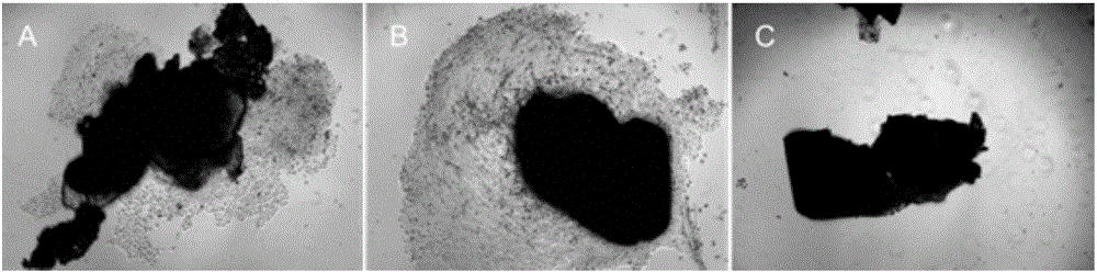
[0033] (1) After fixing the living giant salamander, cut off the skin tissue of about 1 cm × 1 cm in size from its tail, and immediately put it into a pre-cooled 4°C culture medium containing double antibodies (penicillin 500 U / mL, streptomycin 0.5 mg / mL). 60% PBS buffer (dilute the PBS buffer with high-pressure sterilized ultrapure water to a PBS volume fraction of 60%), and transport it to the laboratory ultra-clean workbench within 10 minutes;

[0034] (2) scrape off the mucus on the surface of the giant salamander skin tissue in PBS with ophthalmic tweezers in the ultra-clean workbench, and soak it in 75% alcohol by volume for 10 seconds;

[0035] (3) Wash 6-8 times with 60% PBS buffer solution pre-cooled at 4°C and added with double antibodies (penicillin 500U / mL, streptomycin 0.5mg / mL), and put it into a sterilized ampo...

Embodiment 2

[0043] The method for separating and cultivating giant salamander skin epidermal cells that the present invention adopts comprises the following steps:

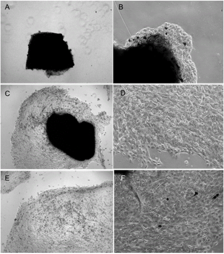
[0044] (1) After fixing the living giant salamander, cut off the skin tissue of about 1 cm × 1 cm in size from its tail, and immediately put it into a pre-cooled 4°C culture medium containing double antibodies (penicillin 500 U / mL, streptomycin 0.5 mg / mL). 60% PBS buffer solution, transported to the laboratory ultra-clean workbench within 10 minutes;

[0045] (2) scrape off the mucus on the surface of the giant salamander skin tissue in PBS with ophthalmic tweezers in the ultra-clean workbench, and soak it in 75% alcohol by volume for 10 seconds;

[0046] (3) Wash 6-8 times with 60% PBS buffer solution pre-cooled at 4°C and added with double antibodies (penicillin 500U / mL, streptomycin 0.5mg / mL), and put it into a sterilized ampoule;

[0047] (4) Use ophthalmic scissors to cut the giant salamander skin tissue into tissue blo...

Embodiment 3

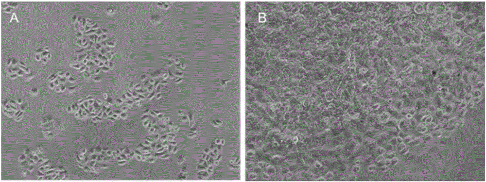
[0057] The present invention uses the giant salamander skin epidermal cells obtained in Example 1 to sequence the giant salamander K5, K10 and P63 genes, including the following steps:

[0058] (1) Primer design

[0059] Since the K5, K10 and P63 gene sequences of giant salamander have not been published yet, we first select species with high homology to giant salamander, and then search the K5, K10 and P63 gene sequences of these species in the NCBI database, and use BioXM software to analyze the gene sequences of these species. K5, K10 and P63 gene sequences were compared, and primers were designed at the parts with high homology. The designed primers are as follows:

[0060] Giant salamander K5 gene upstream primer: 5'-CAGGACTCTGCTTCAACTCG-3'(SEQ ID NO.1); downstream primer: 5'-CGACCAGGAGTAACATTGAAC-3'(SEQ ID NO.2);

[0061] Giant salamander K10 gene upstream primer: 5'-ACTCACCCTGTCCAAATC-3'(SEQ ID NO.3); downstream primer: 5'-TCAGCCATAGCCTCATAC-3'(SEQ ID NO.4);

[0062]...


PUM


Abstract

The invention discloses a method for separating, cultivating and identifying a skin epithelial cell of a giant salamander. According to the method, a skin tissue is acquired from the tail part of the giant salamander, a tissue explant method is used to separate to acquire the primary skin epithelial cell of the giant salamander, the primary skin epithelial cell of the giant salamander is subjected to primary culture and subculture in a specific culture solution (the ingredients comprise DMEM/F12 (60%), 15% of FBS, 5 mug/mL of insulin, and 10 ng/mL of KGF) at the temperature of 28 DEG C, and specific genes (K5, K10 and P63) of the acquired skin epithelial cell of the giant salamander are expressed and identified through RT-PCR (Reverse Transcription-Polymerase Chain Reaction). The homologous design primer of the gene sequence of the related species is used to conduct PCR amplification on the genes K5, K10 and P63 of the giant salamander to acquire the gene sequences of K5, K10 and P63 of the giant salamander for the first time. The provided method for separating, cultivating and identifying the skin epithelial cell of the giant salamander lays foundations for the deep study of the skin of the giant salamander as well as the skin regeneration, trauma repair and tissue engineering skin construction with the help of the skin epithelial cell of the giant salamander.

Description

technical field [0001] The invention belongs to the technical field of cell and tissue engineering, and in particular relates to a method for separating, cultivating and identifying epidermal cells of giant salamander skin. Background technique [0002] The giant salamander is the largest and most precious amphibian in the world. It is a national second-class aquatic wild animal protection. It is also a key development species for agricultural industrialization and characteristic agriculture. It has been cultivated in many places in my country and has formed a certain output. In 2012, the breeding volume of giant salamanders has broken through 3.3 million tails. Studies have shown that the artificially cultured giant salamander skin is rich in type I collagen, accounting for about 62.89% of the total protein in the giant salamander skin. This collagen has similar amino acid composition and properties to mammalian collagen, and has no rejection to the human body. At the same ...

Claims


Application Information

Patent Timeline
IPC IPC(8): C12N5/071C12Q1/68
CPCC12N5/0625C12N2501/117C12N2501/33C12N2509/00C12Q1/6869C12Q2565/125C12Q2531/113
Inventor 郭燕杰杨学义马西亚吴卫妮张延召史明艳张瑞洁
Owner LUOYANG NORMAL UNIV
Why Eureka
Try Eureka