
Frequency domain photoacoustic imaging detection method and system of biological tissues

A technology for biological tissue and photoacoustic imaging, which is used in measurement devices, material analysis by optical means, instruments, etc. Problems such as large weight and volume, to achieve the effect of high safety, improved signal-to-noise ratio, and high imaging contrast

Inactive Publication Date: 2017-05-31
HARBIN INST OF TECH
View PDF5 Cites 6 Cited by
  • Summary
  • Abstract
  • Description
  • Claims
  • Application Information

AI Technical Summary

Problems solved by technology

It has the following disadvantages: (1) The depth resolution of time-domain photoacoustic imaging depends on the bandwidth of the acoustic sensor, and the current technology is difficult to manufacture an acoustic sensor with a large bandwidth and a low signal-to-noise ratio; (2) The jitter of the pulsed laser The error and the transient response error of the acoustic wave sensor are large, so it will adversely affect the inversion accuracy of the absorber structure and physical parameters; (3) The pulsed laser has a high peak power, although its power will be limited to thermal damage However, it may still cause potential adverse effects on biological tissues; (4) The cost of high-power nanosecond pulsed lasers is relatively high, and the weight and volume of equipment are large

Method used


Image

  • Frequency domain photoacoustic imaging detection method and system of biological tissues
  • Frequency domain photoacoustic imaging detection method and system of biological tissues
  • Frequency domain photoacoustic imaging detection method and system of biological tissues

Examples


specific Embodiment approach 1

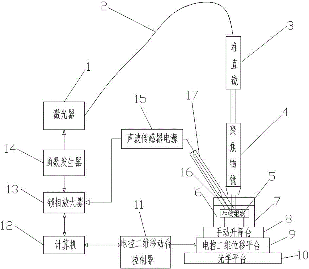
[0029] Specific implementation mode one: as figure 1 Shown, a kind of method for biological tissue frequency-domain photoacoustic imaging detection, described method comprises the following steps:

[0030] Step 1: pre-treat the biological tissue 5 to be measured (i.e., remove hair from the biological tissue 5 to be detected, pre-prepared foreign matter treatment) and make a sample, and place the biological tissue sample into the container 7;

[0031] Step 2: add water 6 into the container 7 as the coupling liquid, immerse the biological tissue sample for 5 mm, place the probe of the liquid-coupled acoustic wave sensor 16 into the water 6 in the container 7, pre-condition the probe of the liquid-coupled acoustic wave sensor 16 Infiltration for 25~35min;

[0032] Step 3: start the computer 12, and run the pre-programmed control software in the computer 12;

[0033] Step 4: Adjust the positions of the collimating lens 3 and the focusing objective lens 4, and adjust the manual l...

specific Embodiment approach 2

[0039] Specific implementation mode two: as figure 1 As shown, a system for realizing the frequency-domain photoacoustic imaging detection method of biological tissue according to claim 1, the system includes a laser 1, an optical fiber 2, a collimating mirror 3, a focusing objective lens 4, water 6, a container 7, a manual Lifting platform 8, electronically controlled two-dimensional displacement platform 9, optical platform 10, electronically controlled two-dimensional displacement platform controller 11, computer 12, lock-in amplifier 13, function generator 14, acoustic wave sensor power supply 15, liquid-coupled acoustic wave sensor 16 and its preamplifier 17;

[0040] Water 6 is added into the container 7 as the coupling liquid, the probe of the liquid-coupled acoustic wave sensor 16 is inserted into the water 6 in the container 7, and the power supply 15 of the acoustic wave sensor is respectively supplied to the liquid-coupled acoustic wave sensor 16 and the front Amplif...

Embodiment 1

[0042] A method for frequency-domain photoacoustic imaging detection of biological tissue, the method comprising the following steps:

[0043] Step 1: pre-treat the biological tissue 5 to be measured (i.e., remove hair from the biological tissue 5 to be detected, pre-prepared foreign matter treatment) and make a sample, and place the biological tissue sample into the container 7;

[0044] Step 2: add water 6 into the container 7 as the coupling liquid, immerse the biological tissue sample for 5 mm, place the probe of the liquid-coupled acoustic wave sensor 16 into the water 6 in the container 7, pre-condition the probe of the liquid-coupled acoustic wave sensor 16 Infiltration for 25 minutes;

[0045] Step 3: start the computer 12, and run the pre-programmed control software in the computer 12;

[0046] Step 4: Adjust the positions of the collimating lens 3 and the focusing objective lens 4, and adjust the manual lifting platform 8 so that the focus of the laser spot is locat...


PUM


Abstract

The invention belongs to the biomedical field, and concretely relates to a frequency domain photoacoustic imaging detection method and system of biological tissues. The method concretely comprises the following steps: 1, preprocessing a biological tissue sample to be detected, and placing the preprocessed sample in a container 7; 2, immersing the sample in water, and pre-infiltrating a liquid coupled sound wave sensor probe in water for 25-35 min; 3, starting a computer to run pre-programming control software; 4, adjusting the spot focus of laser to make the focus positioned on the surface layer of the biological tissue sample; 5, setting the power of a laser device, and setting the parameters of a function generator; 6, running a control program; and 7, running different working modes. The frequency domain photoacoustic imaging detecting method and system of biological tissues overcome the disadvantages of traditional photoacoustic detection methods, and have the advantages of high signal-to-noise ratio, high detection depth, high contrast ratio, low system device cost, good detection effect, simplicity in operation, and convenience in use due to adoption of a frequency domain photoacoustic technology.

Description

technical field [0001] The invention belongs to the field of biomedical nondestructive testing, and in particular relates to a biological tissue frequency-domain photoacoustic imaging detection method and system. Background technique [0002] Biomedical imaging methods are of great significance in clinical diagnosis, experimental research and other fields. Traditional optical imaging methods have been deeply studied and widely used in some biological imaging fields due to their advantages of high resolution and no ionizing radiation. However, due to the scattering effect of biological tissue on light, the imaging depth of traditional optical imaging methods is generally smaller than the mean free path of photons in tissue, and the detection ability of deep biological tissue is low. The photoacoustic imaging method uses scattered photons to excite acoustic signals, and uses acoustic detectors to collect acoustic signals in deep tissues. Since the attenuation of acoustic wave...

Claims


Application Information

Patent Timeline
IPC IPC(8): G01N21/17
CPCG01N21/1702
Inventor 刘俊岩刘洋
Owner HARBIN INST OF TECH
Learn More