

MRNA-coding nanobody and application thereof
A nanobody, coding technology, applied in applications, recombinant DNA technology, antibody mimics/scaffolds, etc., can solve problems such as destruction, cell transformation to produce cancer, and cell function destruction.
- Summary
- Abstract
- Description
- Claims
- Application Information
AI Technical Summary
Problems solved by technology
Method used
Image

Examples
Embodiment 1
[0113] Example 1: Expression and purification of HDAC6-CAT1 truncated protein:
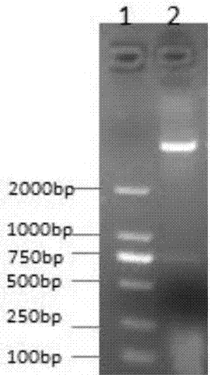
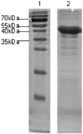
[0114] (1) According to the HDAC6 gene sequence, use Premier Primer5.0 software to design PCR primers: CAT1-JD-5-sal1 (cgaGTCGACgagcagttaaatgaattccattg) and CAT1-JD-3-not1 (gcgGCGGCCGCggcggccatctcacccttggggtcc), the length of the amplified gene fragment is 801bp; ( 2) Using CAT1-JD-5-sal1 and CAT1-JD-3-not1 as primers and HDAC6-WT recombinant plasmid as a template, 6-272 amino acids of HDAC6-Cat1 were amplified. 25 μL of PCR reaction system, containing 12.5 μL of 2× PCR Mix (containing enzyme), 1 μL of upstream primer, 1 μL of downstream primer, 1 μL of DNA template, and 9.5 μL of double distilled water. The cycle parameters of PCR were: pre-denaturation at 95°C for 8min; denaturation at 95°C for 40s, annealing at 57°C for 40s, extension at 72°C for 50s, 35 cycles; final extension at 72°C for 10min. 1% agarose gel electrophoresis observation. (3) Construct the pET32a-Cat1-JD recombinant plasmid,...
Embodiment 2
[0115] Example 2: Construction of the HDAC6-CAT1 nanobody library:
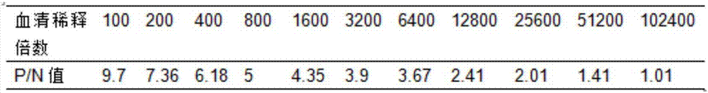
[0116] (1) Mix 1 mg of CAT1-JD recombinant protein antigen with Freund's adjuvant in equal volumes, and immunize a Xinjiang Bactrian camel once a week for a total of 7 consecutive immunizations. During the immunization process, B cells are stimulated to express specific nanobodies (2) After 7 times of immunization, extract 100ml of camel peripheral blood lymphocytes, and carry out ELISA antibody level detection of immunized camels, the results are as follows: image 3 As shown, the serum titer after immunization reached 10 4 , indicating that the HDAC6-CAT1-JD recombinant protein has a better immune effect; and extract total RNA; (3) synthesize cDNA and amplify VHH by nested PCR; (4) digest 20ug pMECS phage with restriction enzymes PstⅠ and NotⅠ Display the vector and 10ug VHH and connect the two fragments; (5) Transform the ligation product into electroporation competent cells TG1, construct the human antib...
Embodiment 3
[0117] Embodiment 3: Nanobody screening process against CAT1-JD recombinant protein:
[0118] (1) Take 200uL of recombinant TG1 cells and culture them in 2×TY medium, add 40uL of helper phage VCSM13 to infect TG1 cells during the period, and culture overnight to amplify the phages, use PEG / NaCl to precipitate the phages the next day, and centrifuge to collect the amplified phages ; (2) Couple 200ug of CAT1-JD recombinant protein dissolved in 100mM pH 8.2 NaHCO3 to a microtiter plate, place it overnight at 4°C, and set up a negative control at the same time; (3) Add 100ul of 3% BSA the next day, Block at room temperature for 2h; (4) After 2h, add 100ul amplified phage (2×10 11 tfu immunized camel nanobody phage display gene library), and reacted at room temperature for 1h; (5) Washed 5 times with PBS+0.05%Tween-20 to wash off the bound phage; (6) Use trypsin with a final concentration of 25mg / ml The phage specifically bound to the Fc fragment of the human antibody will be diss...
PUM
 Abstract
Description
Claims
Application Information