Intravascular virtual endoscope imaging system based on electromagnetic positioning composite conduit and working method of system

A virtual endoscope and electromagnetic positioning technology, applied in the field of intravascular interventional imaging and diagnosis, can solve the problems of complex operation, inability to realize deep blood vessels, and inability to provide endoscopic images, etc.

Active Publication Date: 2017-08-08
SHANDONG UNIV QILU HOSPITAL +1
View PDF10 Cites 12 Cited by
  • Summary
  • Abstract
  • Description
  • Claims
  • Application Information

AI Technical Summary

Problems solved by technology

However, this patent has the following defects: the body surface three-dimensional ultrasound instrument used in the method described in this patent can only obtain images of superficial organs, and cannot realize imaging of deep blood vessels, such as coronary arteries and intracranial blood vessels, and cannot provide endoscopic imaging. In addition, CT or MRI and interventional catheters are needed to obtain the vascular skeleton first, then perform ultrasound imaging, and finally realize multi-step and multi-modal imaging of image matching
The actual operation of this patent needs to be carried out separately, that is, the use of body surface ultrasound for 3D reconstruction before operation, and the use of interventional catheters to obtain and match the path during the operation, which is complicated.

Method used


Image

  • Intravascular virtual endoscope imaging system based on electromagnetic positioning composite conduit and working method of system
  • Intravascular virtual endoscope imaging system based on electromagnetic positioning composite conduit and working method of system
  • Intravascular virtual endoscope imaging system based on electromagnetic positioning composite conduit and working method of system

Examples


Embodiment 1

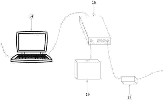
[0068] The intravascular virtual endoscopic imaging system based on the electromagnetic positioning composite catheter includes an electromagnetic positioning tracking module, a composite intravascular imaging catheter module, a positioning information processing and composite image presentation module 14, an electromagnetic positioning tracking module, and a composite intravascular imaging catheter module. Connect the positioning information processing and composite image presentation module 14;

[0069] The composite intravascular imaging catheter module is used to obtain intravascular images, and the electromagnetic positioning tracking module is used to obtain the movement track of the composite intravascular imaging catheter module in the blood vessel, and transmit it to the positioning information processing and composite image presentation module 14 to perform intravascular images Correction fusion with positioning information presents vessel spatial information in a vir...

Embodiment 2

[0072] According to the intravascular virtual endoscopic imaging system based on the electromagnetic positioning composite catheter described in Embodiment 1, the difference is that, as image 3 As shown, the composite intravascular imaging catheter module includes a sensor coil unit 6, an intravascular imaging probe unit 8, and a stepping motor unit. The sensor coil unit 6 and the intravascular imaging probe unit 8 are connected in parallel, and the sensor is driven by the stepping motor unit. The coil unit 6 and the intravascular imaging probe unit 8 advance and retreat synchronously; realize the anti-bending, flexibility and pushability of the catheter in the blood vessel;

[0073] The intravascular imaging probe unit 8 is an intravascular imaging catheter, and the top end of the intravascular imaging catheter is provided with an intravascular imaging probe 12;

[0074] The sensor coil unit 6 is used to obtain the position information of 5 degrees of freedom, including the ...

Embodiment 3

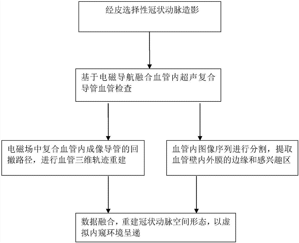
[0093] The working method of the intravascular virtual endoscopic imaging system based on the electromagnetic positioning composite catheter described in embodiment 2, such as figure 2 As shown, this embodiment is described by taking the rabbit abdominal aorta examination as an example, including:

[0094] (1) The femoral artery is successfully punctured, the sheath is placed, and the guide wire 10 is inserted retrogradely through the guide wire through the outlet 7 to reach the junction of the thoracic and abdominal aorta, and the intravascular imaging probe unit is flushed through the cavity interface 3 of the outer packaging tube 8. The intravascular imaging probe unit 8 of this embodiment is an intravascular ultrasound catheter, and the composite intravascular imaging catheter is sent along the guide wire 10 according to the position of the X-ray-opaque marker 13 at the head end of the composite catheter under X-ray Reach the abdominal aorta, fix the annular bulge 4 to th...


PUM


Abstract

The invention relates to an intravascular virtual endoscope imaging system based on an electromagnetic positioning composite conduit and a working method of the system. The intravascular virtual endoscope imaging system comprises an electromagnetic positioning tracking module, a composite intravascular imaging conduit module and a positioning information processing and composite image presenting module, wherein the composite intravascular imaging conduit module is used for acquiring intravascular images; the electromagnetic positioning tracking module is used for acquiring a movement track of the composite intravascular imaging conduit module inside blood vessels, and used for transmitting the movement track to the positioning information processing and composite image presenting module, then correction and fusion of the intravascular images and the positioning information is implemented, and blood vessel spatial information is represented in a virtual endoscope environment. Relatively precise bases are provided for diagnosis on vasculopathy.

Description

technical field [0001] The invention relates to an intravascular virtual endoscope imaging system based on an electromagnetic positioning composite catheter and a working method thereof, belonging to the technical field of intravascular interventional imaging and diagnosis. Background technique [0002] According to my country's 2008 China Health Care Development Statistical Bulletin, cardiovascular disease is also the number one cause of death among urban and rural residents in my country, with an average of one person dying every 10 seconds, which is three times the death rate of cardiovascular disease in the United States. (1 death every 34 seconds)! The high morbidity and mortality of cardiovascular diseases have become an important factor restricting the social and economic development of our country. Recent studies have found that in most patients with acute cardiovascular and cerebrovascular events, atherosclerotic (AS) plaque rupture and thrombosis are the main patho...

Claims


Application Information

Patent Timeline
Patent Type & Authority Applications(China)
IPC IPC(8): A61B5/06A61B8/08A61B8/12
CPCA61B5/0035A61B5/0075A61B5/0084A61B5/0095A61B5/062A61B5/6852A61B5/7225A61B8/0891A61B8/12A61B8/469A61B8/485A61B8/52A61B8/5261
Inventor 张鹏飞苏海军冯建钦丁毅寿张昱昕
Owner SHANDONG UNIV QILU HOSPITAL
Why Eureka
Try Eureka