

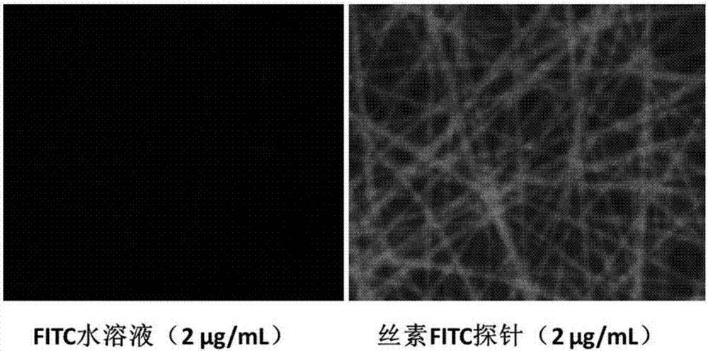
Preparation method of fibroin fluorescent probe
A fluorescent probe and silk fibroin technology, which is applied in the biological field and achieves the effects of wide application prospects, high sensitivity and convenient sample preparation process.
- Summary
- Abstract
- Description
- Claims
- Application Information
AI Technical Summary
Problems solved by technology
Method used
Image

Examples
Embodiment 1
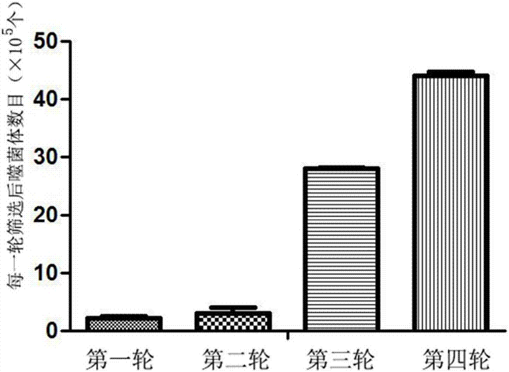
[0046] 1) Use the phage peptide library produced by commercial NEB to carry out 5 rounds of screening on the silk membrane to obtain the phage bound to silk fibroin, the specific process is as follows;
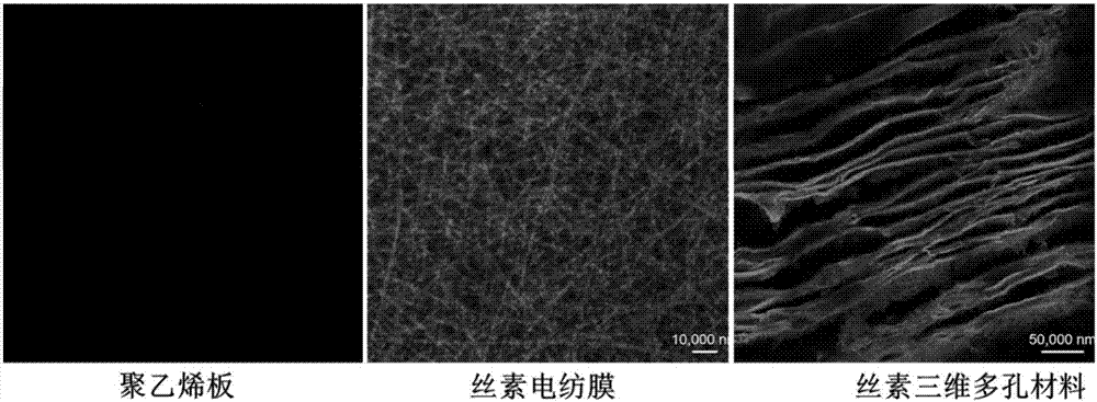
[0047] 1.1) Preparation of silk film:
[0048]a. Obtain an aqueous silk protein solution with a concentration of 1 mg / mL;
[0049] b. Drop the fibroin solution on a polyethylene plate to air-dry, add alcohol to make it solidify, and obtain a water-insoluble silkworm fibroin film.
[0050] 1.2) Screening of polypeptide sequences bound to silk fibroin
[0051] a. Sealed silk fibroin material: add the buffer solution of TBS+BSA (0.5mg / ml) to the silkworm silk fibroin film;
[0052] b. Screening of phage library by silk fibroin material: For each phage species, add 10 microliters of the commercialized M13-12 phage random display library to the silk film and shake at 150rpm for 0.5h in a shaker at room temperature;
[0053] c. Washing: wash the silk fibroin film once with TBST w...
Embodiment 2
[0065] 1) Use the phage peptide library produced by commercial NEB to carry out 5 rounds of screening on the silk membrane to obtain the phage bound to silk fibroin, the specific process is as follows;
[0066] 1.1) Preparation of silk film:
[0067] a. Obtain an aqueous silk protein solution with a concentration of 10 mg / mL;
[0068] b. Drop the fibroin solution on a polyethylene plate to air-dry, add alcohol to make it solidify, and obtain a water-insoluble silkworm fibroin film.
[0069] 1.2) Screening of polypeptide sequences bound to silk fibroin
[0070] a. seal silk fibroin material: add the buffer solution of TBS+BSA (10mg / ml) in silkworm silk fibroin film;
[0071] b. Screening of phage library by silk fibroin material: for each phage species, 10 microliters of commercial M13-12 phage random display library was added to the silk film, and shaken at 220rpm for 0.5h in a shaker at room temperature;
[0072] c. Washing: wash the silk fibroin film 10 times with TBST wa...
Embodiment 3
[0084] 1) Use the phage peptide library produced by commercial NEB to carry out 5 rounds of screening on the silk membrane to obtain the phage bound to silk fibroin, the specific process is as follows;
[0085] 1.1) Preparation of silk film:
[0086] a. obtain silk protein aqueous solution, concentration is 50mg / mL;
[0087] b. Drop the fibroin solution on a polyethylene plate to air-dry, add alcohol to make it solidify, and obtain a water-insoluble silkworm fibroin film.
[0088] 1.2) Screening of polypeptide sequences bound to silk fibroin
[0089] a. Sealed silk fibroin material: add the buffer solution of TBS+BSA (1mg / ml) to the silkworm silk fibroin film;
[0090] b. Screening of phage library by silk fibroin material: for each phage species, 10 microliters of commercial M13-12 phage random display library was added to the silk film, and shaken at 220rpm for 0.5h in a shaker at room temperature;
[0091] c. Washing: wash the silk fibroin film 10 times with TBST washing...
PUM
| Property | Measurement | Unit |
|---|---|---|
| molecular weight | aaaaa | aaaaa |
Abstract
Description
Claims
Application Information