HN-VP233-221aa fusion protein as well as preparation method and application thereof

A technology of fusion protein and bursa, which is applied in the field of HN-VP233-221aa fusion protein and its preparation, can solve the problems of reduced immune effect of traditional vaccines, increased susceptibility to other diseases, impaired lymphocyte function, etc., and achieve enhanced body fluid and cellular immune response, good immune protection rate, good protective effect

Active Publication Date: 2017-10-13
HENAN UNIV OF SCI & TECH
4 Cites 0 Cited by

AI-Extracted Technical Summary

Problems solved by technology

IBD mainly affects chicks and young chickens aged 3-6 weeks, and can destroy the central organ of humoral immunity in chickens—the bursa of Fabricius, resulting in impaired lymphocyte function and immunosuppression, thereby affecting the immune effects of vaccines such as Newcastle disease and avian influenza. and increased susceptibility to other diseases
At present, due to the pre...

Method used

HN-VP233-221aa fusion protein gene is formed by the tandem connection of flexible Linker gene by chicken infectious bursal virus VP2 main protective antigen fragment (97-663bp) and chicken Newcastle disease virus HN gene, the tandem dna sequence The encoded amino acid sequence is the HN-VP233-221aa fusion protein. The fusion protein can induce chicks to produce high levels of HN-specific antibo...

Abstract

The invention discloses HN-VP233-221aa fusion protein as well as a preparation method and application thereof. The HN-VP233-221aa fusion protein comprises an amino acid sequence of infectious bursal disease virus VP233-221aa, an amino acid sequence of Newcastle disease virus hemagglutinin-neuraminidase (HN protein) and a flexible Linker peptide amino acid sequence located between the amino acid sequence of the VP233-221aa and the amino acid sequence of the HN protein of Newcastle disease virus; the HN-VP233-221aa fusion protein is formed by connecting a main protective gene segment of VP2 and an HN gene of the Newcastle disease virus in series through a flexible Linker peptide (-G-S-). The recombinant HN-VP233-221aa fusion protein disclosed by the invention can be used for effectively inducing an organism to generate body fluid and cell immune response and can be used for inducing immunized chickens to generate high-level HN specific antibodies and VP2 antibodies and accelerating a proliferation and activation capability of chicken T lymphocytes; the HN-VP233-221aa fusion protein has a very good protection effect on attacks of infectious bursal disease virulent strains and Newcastle disease virulent strains so that the effect of preventing two diseases through one injection is realized.

Application Domain

SsRNA viruses negative-senseViral antigen ingredients +10

Technology Topic

Cell immune responseT lymphocyte +16

Image

  • HN-VP233-221aa fusion protein as well as preparation method and application thereof
  • HN-VP233-221aa fusion protein as well as preparation method and application thereof
  • HN-VP233-221aa fusion protein as well as preparation method and application thereof

Examples

  • Experimental program(3)

Example Embodiment

[0049] Recombinant HN-VP2 of the present invention 33-221aa The preparation method of the fusion protein includes the following steps:
[0050] (A) HN-VP2 33-221aa And HN gene sequence acquisition
[0051] ①IBDV VP2 33-221aa Obtaining the gene sequence:
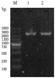
[0052] According to the nucleotide sequence of IBDV VP2 in GenBank (No:AF508177), design a pair of primers to amplify VP2 33-221aa Gene fragment.
[0053] The pMD18-T-VP2 constructed in the previous stage was used as a template, and IBDV VP2 was amplified by PCR 33-221aa Fragment. After the amplified product was identified by 1% agarose gel electrophoresis, the target band was cut out and recovered and purified using a common agarose gel DNA recovery kit. The purified product is VP2 33-221aa Gene fragments and send them to Dalian Bao Biological Engineering Co., Ltd. for sequencing;
[0054] ②Acquisition of NDV HN gene
[0055] According to the nucleotide sequence of NDV HN gene in GenBank (No: JF343538), a pair of primers was designed to amplify the HN gene fragment of Newcastle disease virus.
[0056] The NDV strain was inoculated with 9-10 day-old SPF chicken embryos through the allantoic cavity route, and the allantoic fluid of the chicken embryos was collected and stored at -20°C until use. Follow the instructions of Dalian Bao Biological Engineering Co., Ltd. RNA extraction kit to extract NDV RNA template. And use total RNA as template for RT-PCR. After the PCR product was identified by 1% agarose gel electrophoresis, the target band was cut, and the target gene was recovered and purified using a DNA recovery kit. The purified product was the Newcastle disease virus HN gene and sent to Dalian Bao Bioengineering Co., Ltd. for sequencing;
[0057] (B) HN-VP2 33-221aa Obtaining fusion gene sequence
[0058] VP2 obtained by ①② in (a) 33-221aa The gene fragment and the HN gene of Newcastle disease virus were used as templates, and HN-VP2 was amplified by the overlap extension PCR method 33-221aa Fusion gene.
[0059] After the PCR product was identified by electrophoresis on a 1% agarose gel containing ethidium bromide, the target band was cut, and then the HN-VP2 was recovered according to the instructions of the gel recovery kit. 33-221aa Fusion gene fragments, and sent to Dalian Bao Biological Engineering Co., Ltd. for sequencing.
[0060] (C) Highly express HN-VP2 33-221aa Construction of genetically engineered strain of fusion protein
[0061] The HN-VP2 obtained above 33-221aa The fusion protein gene fragment was inserted into the prokaryotic expression vector pET32a to obtain the recombinant vector pET32a-HN-VP2 33-221aa; Then the recombinant plasmid was transformed into Rosetta Escherichia coli, through resistance screening, PCR amplification and restriction enzyme digestion of suspected colonies were carried out to obtain Rosetta expressing recombinant fusion protein (pET32a-HN-VP2 33-221aa ) Genetically engineered strains;
[0062] (D) Soluble HN-VP2 33-221aa Acquisition of fusion protein
[0063] Will identify the correct Rosetta (pET32a-HN-VP2 33-221aa The single colony of) was inoculated into LB liquid medium and cultured overnight at 37°C. The cultured bacterial solution was transferred to LB liquid medium at a ratio of 1:100, and cultivated at 37°C until it became cloudy, and IPTG was 1:1: Add in the ratio of 50, induce culture for 6 h, centrifuge at 5000r/min for 10 min to collect the culture supernatant, which is the obtained HN-VP2 33-221aa Fusion protein.

Example Embodiment

[0065] Example 1
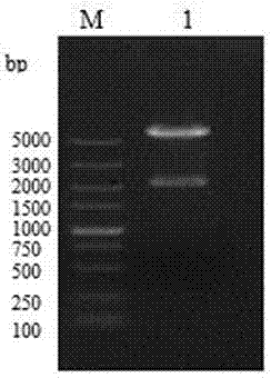
[0066] 1. HN-VP2 33-221aa Acquisition of fusion protein coding gene sequence
[0067] (1) Acquisition of HN gene of Newcastle disease virus
[0068] According to the nucleotide sequence of the HN gene of Newcastle disease virus in GenBank (No: JF343538), a pair of specific primers were designed to amplify the HN gene fragment of Newcastle disease virus using DNA Star7.0 and Primer premier 6.0 software.
[0069] P1 and P2 amplify the HN gene of Newcastle disease virus, and introduce the 5'end of primer P1 Bam At the HI endonuclease site, a flexible linker was introduced at the 5'end of P2. The primer sequence is as follows:
[0070] Table 1 The primer sequences used in PCR amplification
[0071]
[0072] The frozen NDV strains were thawed in a 37°C water bath and then inoculated with 9-10 day-old non-immunized chicken embryos (0.2 mL per embryo) through the allantoic cavity, and incubated at 37°C. Collect aseptically the allantoic fluid of dead chicken embryos for 24 to 48 hours (the chicken embryo fluid that has red blood cells or egg yolk makes the allantoic fluid turbid is discarded), after repeated freezing and thawing 3 times, centrifuged at 8 000r/min 4℃ for 15min, Take the supernatant and store at -20°C until use. The extraction of viral RNA is performed in accordance with the instructions of the RNA extraction kit. Using 4 μL of extracted total RNA as a template, add MgCl 2 3 μL, 10 × reverse transcription buffer 2.5 μL, dNTP Mixture (10 mmol/L) 2.5 μL, RNaseInhititor 0.5 μL (20U), RT enzyme 0.5 μL, Taq Enzyme 0.5 μL, primer P1 1 μL, primer P2 1 μL, sterilized dH 2 O9.5 μL, total system 25 μL. Perform the RT-PCR reaction under the following reaction conditions: 50 ℃ 40 s, 94 ℃ 2 min, 94 ℃ 45 s, 53 ℃ 90 s, 72 ℃ 100 s, cycle 30 times, and finally 72 ℃ extension 10 min. After the PCR product was identified by electrophoresis on a 1% agarose gel containing ethidium bromide, the target band was cut, and then recovered according to the gel recovery kit instructions to be the Newcastle disease virus HN gene.
[0073] (2) VP2 33-221aa Acquisition of gene sequence
[0074] According to the IBDV VP2 nucleotide sequence in GenBank (No:AF508177), a pair of specific primers P3 and P4 were designed to amplify VP2 using DNA Star7.0 and Primerpremier6.0 software 33-221aa. The primers were synthesized by Dalian Bao Biological Engineering Company. Primer P3 5’ end is introduced into the flexible joint, P4 5’ end Hin dⅢ endonuclease site. The primer sequence is as follows:
[0075] Table 2 The primer sequences used in PCR amplification
[0076]
[0077] The pMD18-T-VP2 constructed in the previous stage was used as a template to amplify chicken VP2 by PCR 33-221aa. Add 10×PCR Buffer, 5 μL, MgCl to the reaction system 2 , 3 μL; dNTP, 2.5 mmol/L, 4 μL; pMD18-T-VP2 template 0.5 μL, primer P3 and primer P4, the final concentration is 10 pmol/L each 2 μL; TaKaRa Ex Taq 0.5 μL; sterilized ultrapure water, 33 μL; reaction conditions: 95 ℃ pre-denaturation 5 min; 95 ℃ denaturation 30 s; 63 ℃ annealing for 30 s; 72 ℃ extension 40 s, 30 cycles; 72 ℃ extension 10 min . After the PCR reaction is over, perform agarose gel electrophoresis and observe the results. After the PCR amplification product was identified by 1% agarose gel electrophoresis, the target band was cut out and recovered and purified using the ordinary agarose gel DNA recovery kit of Dalian TaKaRa Company. The purified product is VP2 33-221aa The gene fragments will be sent to Dalian Biotech for sequencing.
[0078] (3) HN-VP2 33-221aa Acquisition of fusion protein coding gene sequence
[0079] Use the above recovered and purified HN/VP2 gene fragment as a template, use P1/P4 primers, and obtain HN-VP2 by SOEing PCR technology 33-221aa Fusion fragments.
[0080] PCR reaction system 50 μL: 10×PCR Buffer, 5 μL, MgCl 2 , 3 μL; dNTP, 10 mmol/L, 1 μL; VP2 33-221aa And Newcastle disease virus HN gene as templates, add 5 μL each, primer P1 and primer P4, the final concentration is 20 pmol/L each 2 μL; TaKaRa Ex Taq 0.5 μL; sterilized ultrapure water, 34.5 μL;
[0081] PCR reaction conditions: 95 ℃ pre-denaturation for 5 min; 95 ℃ denaturation for 30 s; 63 ℃ annealing for 30 s; 72 ℃ extension for 2 min 20 s, 30 cycles; 72 ℃ extension for 10 min, PCR product containing 1% ethidium bromide After identification by agarose gel electrophoresis, the target band is cut out, and then recovered according to the instructions of the gel recovery kit, which is the HN-VP2 fusion protein encoding gene (SEQ.ID.NO.5).
[0082] 2. Recombinant plasmid pET32a-HN-VP2 33-221aa Construction and identification of
[0083] First, HN-VP2 33-221aa Link to pMD19-T vector, and then use Bam HⅠ, Hin dⅢ to pMD19-T-HN-VP2 33-221aa Carry out double enzyme digestion and recover the fusion gene fragment HN-VP2 33-221aa. At the same time use pET32a Bam HⅠ, Hin dⅢ Perform double digestion and recover the vector fragment. The recovered HN-VP2 33-221aa Connect with pET32a, ligation system 10 µL: target fragment 6 µL, vector 2 µL, T4 DNA ligase 1 µL, Buffer 1 µL. The connection conditions are 12-16°C overnight. Mix the Rosetta competent cells and the ligation product in a test tube in an ice bath for 30 min, heat shock at 42°C for 90 s, and ice bath for 2 min. Add 400 µL of LB medium to the test tube, and incubate in a constant temperature shaker at 37°C for 1.5 h , Take 100 µL for plating (LB+AMP), incubate at 37°C for 10-12 hours, pick a single colony for PCR and double enzyme digestion identification, such as Figure 1-2 , The plasmid with a vector band of about 5900bp and a DNA fragment of 2300bp after digestion was used as the positive plasmid, and the positive plasmid was named pET32a-HN-VP2 33-221aa , Sent to Shanghai Invitrongen company for sequencing.
[0084] Three, express HN-VP2 33-221aa Construction of genetically engineered strains of fusion protein:
[0085] Take the positive recombinant expression vector pET32a-HN-VP2 obtained above 33-221aa Mix 5 μg with 80 μl LRosetta competent cells, mix well in a test tube and ice bath for 30 min, then heat shock at 42°C for 90 s, then ice bath for 2 min, finally add 400 μL LB medium to the test tube, culture for 1.5 h, then Coat the plate with 100 µL (LB+AMP), culture for 10~12 h, pick a single colony for PCR identification, the reaction system is the same as above, PCR reaction conditions: 95℃ pre-denaturation for 5 min; 95℃ denaturation for 30 s; 63℃ annealing for 30 s; 72 ℃ extension for 2 min 20 s, 30 cycles; 72 ℃ extension for 10 min, amplify a 2300bp gene fragment, and transfer the identified single colony to LB+AMP liquid medium, extract the positive plasmid, and then use Bam HⅠ, Hin dⅢ Double enzyme digestion identification, the digestion system is 10×H buffer 1 µL, Bam HⅠ 0.4 µL, Hin dⅢ 0.4 µL, recombinant plasmid 8.2 µL; digestion conditions are all 37ºC constant temperature water bath for 3 hours. PCR and restriction enzyme digestion identify the correct colony as expressing HN-VP2 33-221aa A genetically engineered E. coli strain for the fusion protein.
[0086] Fourth, reorganize HN-VP2 33-221aa Induced expression and identification of fusion protein:
[0087] Will identify the correct Rosetta (pET32a-HN-VP2 33-221aa ) A single colony is transferred to the LB+AMP liquid medium and cultured for 10-12 hours, and the cultured bacterial liquid and medium are expanded and cultured for about 5 hours after 1:100. Add 80 µL of inducer IPTG (the ratio of inducer to bacterial solution is 1:50) and shake for 6 h. After that, take out the bacterial solution, put the induced bacteria into a 1.5 mL EP tube, centrifuge, remove the supernatant, add PBS or 200 µL of ultrapure water to wash 2~3 times, and finally add 200 µL of ultrapure water to disrupt the cells with ultrasound The instrument is broken until the solution is transparent. Then add 50 µL of 5× sample solution (the ratio of sample solution to solution is 1:4) into the solution, boil it in boiling water for 10 minutes, and mark it.
[0088] Prepare 5 mL separation gel, use a 5 mL pipette to take an appropriate amount into the gel plate, add an appropriate amount of isobutanol to eliminate bubbles, and let it stand for about 1 h until the separation gel is solidified; use filter paper to completely suck out the isobutanol and prepare 2 mL concentrate Add the gel to the gel plate, quickly insert the comb, and let it stand for 1 hour for the gel to solidify; place the gel plate in the appropriate position in the electrophoresis tank, add appropriate buffer, pull out the comb for spotting, connect the power supply, and concentrate the gel with constant pressure Run at 80 V for about 45 minutes, wait until the bromophenol blue runs through the concentrated gel, and then keep the pressure at 120 V until some of the bromophenol blue runs out of the buffer in the sample, and disconnect the power supply.
[0089] Take out the electrophoresis gel, stain it with Coomassie Brilliant Blue for 1 h, then decolorize it with a decolorizing solution, change it every 2 h, and repeat about 4 times. Analyze protein expression in a gel imager. The recombinant HN-VP233-221aa fusion protein has a molecular weight of about 104 kDa, such as image 3.
[0090] At the same time, the NDV positive serum was used as the primary antibody for Western blotting. The lane where the recombinant E. coli culture solution was loaded had a specific band of interest, while the lane where the negative control was loaded did not appear. Figure 4. It shows that the recombinant HN-VP233-221aa fusion protein has been successfully expressed and has good immunoreactivity.

Example Embodiment

[0091] Example 2
[0092] Recombinant HN-VP2 33-221aa Application of fusion protein:
[0093] 1) Animal immunity
[0094] Use the above recombinant HN-VP2 33-221aa The fusion protein was used to immunize chickens and evaluate their immune characteristics. 240 14-day-old healthy chicks were randomly divided into 4 groups, 60 per group. The first group was a negative control and was immunized with 200 μL PBS; the second group was immunized with 200 μg of HN-VP2 recombinant fusion protein (diluted with 200 μL PBS), and immunized by chest intramuscular injection. Groups 3 and 4 were immunized with Newcastle disease IV vaccine and bursal disease vaccine (B87 strain) as the traditional vaccine control group. 200 μL was immunized by nasal drops and eye drops; all experimental groups were immunized once every 2 weeks. A total of 3 immunizations. And on the 7th, 21st, and 35th day after the first immunization, 8 chicks were randomly selected from each group for cardiac blood collection and serum separation. At the same time, aseptically take the spleen for lymphocyte proliferation test. Two weeks after the last immunization, 18 test chickens from each group were challenged with NDV F48E9 standard virulent virus (1000 ELD 50 /Only), the remaining 18 will be challenged with IBD standard poison;
[0095] After the challenge, observe for 7 consecutive days, and record the morbidity and death of the experimental chickens in each group.
[0096] 2) Determination of NDV HN antibody and IBDV VP2 antibody
[0097] The serum was separated at 7, 21, and 35 days after the first immunization, and the production of HN antibody and VP2IgG antibody in each group of experimental chickens after immunization was detected by ELISA, and the recombinant fusion protein HN-VP2 was found 33-221aa NDV-HN and IBDV-VP2 antibodies can be detected in the immunization group one week after the first immunization. After the second booster immunization, the antibody production continues to increase, and the antibody levels are equivalent to those of the traditional vaccine group, with no significant difference ( Figure 5-6 ).
[0098] 3) Analysis by tetramethylazazole blue method (MTT)
[0099] From the 7th, 21st, and 35th days after the first immunization, the spleens of each test chicken were aseptically collected to prepare a single cell suspension to detect the proliferation of splenic T lymphocytes. The results show different detection points HN-VP2 after immunization 33-221aa The fusion protein immunization group has the strongest ability to induce the proliferation of T lymphocytes, and there are significant differences between it and other groups in the same period ( p <0.05) ( Figure 7-8 ).
[0100] 4) Determination of IL-4 and IFN-γ in serum
[0101] Quantitative analysis of IL-4 and IFN-γ in serum using quantitative ELISA found that HN-VP2 33-221aa The fusion protein immune group induced lymphocytes to secrete the strongest levels of specific IL-4 and IFN-γ, with significant differences ( p <0.05). Followed by the Newcastle disease IV vaccine group and the infectious bursal disease vaccine group ( p <0.05) ( Figure 9-10 ).
[0102] 5) Challenge test results
[0103] Two weeks after the last immunization, the remaining 18 chickens in each immunization group were treated with F48E9 virulent challenge and IBD standard virulent challenge. The results are shown in Table 3-4. The situation of NDV virulent challenge: In the HN-VP2 immunization group, 18 chickens in the HN-VP2 immunization group had 2 cases of disease during the 7-day observation period, of which 1 died, with a protection rate of 88.9%; and IV There was 1 animal in the vaccine group, and the protection rate was 94.4%. IBDV standard violent poisoning situation: HN-VP2 33-221aa Three of the 18 experimental chickens in the fusion protein immunization group developed disease during the 7-day observation period, with a protection rate of 83.3%. The protection rate of the traditional vaccine group was 88.9% (Table 1).
[0104] Table 3 The protection rate of each group after NDV challenge
[0105]
[0106] Table 4 The protection rate of each group after IBDV challenge
[0107]

PUM

PropertyMeasurementUnit
Molecular weight104.0

Description & Claims & Application Information

We can also present the details of the Description, Claims and Application information to help users get a comprehensive understanding of the technical details of the patent, such as background art, summary of invention, brief description of drawings, description of embodiments, and other original content. On the other hand, users can also determine the specific scope of protection of the technology through the list of claims; as well as understand the changes in the life cycle of the technology with the presentation of the patent timeline. Login to view more.

Similar technology patents

Waterproof safe socket

Owner:应城御尚智能家居设计有限公司

Wheel bearing arrangement comprising an encoder and a sensor

InactiveUS7618191B2good protection
Owner:SCHAEFFLER TECH AG & CO KG

Longitudinal tear detection device and method for conveying belt

Owner:河北省机电一体化中试基地有限公司
Why Eureka
Try Eureka