Fluorescence quantitative in-situ hybridization (Q-FISH) method for determination of telomere length by using genomic DNA

A technology for telomere length and fluorescence quantification, which can be used in recombinant DNA technology, DNA/RNA fragments, and microorganism determination/inspection. , the effect of improving the accuracy

Active Publication Date: 2018-05-08
济南海湾生物工程有限公司
View PDF4 Cites 5 Cited by
  • Summary
  • Abstract
  • Description
  • Claims
  • Application Information

AI Technical Summary

Problems solved by technology

[0012] Aiming at the problems in the prior art that only the average telomere length can be measured and the variation of telomere length cannot be measured at the same time, the present invention provides a quantitative fluorescence in situ hybridization (Q-FISH) me

Method used


Image

  • Fluorescence quantitative in-situ hybridization (Q-FISH) method for determination of telomere length by using genomic DNA
  • Fluorescence quantitative in-situ hybridization (Q-FISH) method for determination of telomere length by using genomic DNA
  • Fluorescence quantitative in-situ hybridization (Q-FISH) method for determination of telomere length by using genomic DNA

Examples


Embodiment 1

[0066] Example 1 Preparation and effects of telomere-specific PNA probes.

[0067] 1.1 Preparation of telomere length standard

[0068] The base sequence of the telomere-specific PNA probe is shown in SEQ ID NO:1, the N-terminal is modified with a fluorescent group Cy3, and an O-linker is added between the fluorescent group and the N-terminal base, and its sequence is: Cy3- O-TT A GGG TTAGGG, bases underlined are modified miniPEGs, synthesized by PNA Bio (Thousands Oaks, CA).

[0069] 1.2 Fluorescent detection of telomere-specific PNA probes binding to telomeres

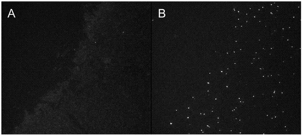
[0070] Two slides loaded with the same DNA samples extracted from blood leukocytes were treated with 0.3 M YoYo-1 dye and telomere-specific PNA probes, and fluorescent images were taken under a 100X oil microscope (Leica DM400B Fluorescence microscope connected to Hamamatsu ORCA-flash 2.8 digital camera, exposure 250 ms, Metamorph image acquisition software), picture as figure 1 Shown: figure 1 A is an image ...

Embodiment 2

[0071] Example 2 Preparation of Telomere Length Standards.

[0072] 2.1 Preparation of telomere length standard

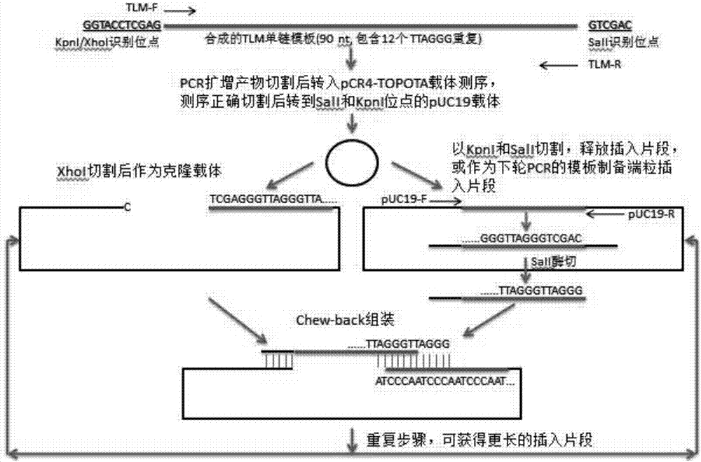
[0073] The preparation process of telomere length standard is as follows: figure 2 , the specific process is as follows:

[0074] (1) Synthesize single-stranded telomeric repeat fragments, which contain 12 telomeric repeat sequences, and the 5' end contains KpnI and wxya site, the 3' end contains SalI site, its sequence is shown in SEQ ID NO: 2, synthesized by Integrated DNA Technologies, Inc;

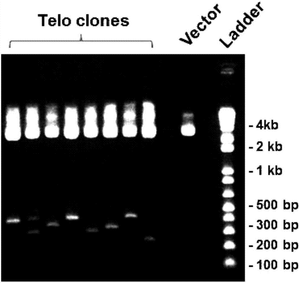
[0075] (2) Perform PCR using the above telomeric repeat fragment as a template. The sequence of the forward primer (TLM-F) of PCR is shown in SEQ ID NO: 3, and the sequence of the reverse primer (TLM-R) is shown in SEQ ID NO: 4 Annealed at 55 degrees, amplified for 30 cycles to obtain PCR products; the PCR products were cloned into pCR4-TOPO TA vector (Life Technologies, Grand Island, NY), and verified by Sanger DNA sequencing (Genwiz, Germantown, MD) sequence is c...

Embodiment 3

[0085] Example 3 Determination of telomere length using the Q-FISH method of genomic DNA.

[0086] 3.1 Determination of Telomere Length Using Genomic DNA Q-FISH

[0087] (1) Genomic DNA was extracted from primary human embryonic lung fibroblasts (WI38) and human breast cancer cells (MCF-7).

[0088] (2) 100 ng of genomic DNA extracted from the two cell lines and telomere length standards of different lengths (100 bp, 200 bp, 400 bp, 600 bp, 900 bp, 1.2 kb, 2.4 kb, 3.6 kb) were coated separately Heating at 75°C for 1 hour on a slide area with a diameter of 0.5 cm, then fixing in cold methanol at 0-4°C, and drying the slide to obtain a slide covered with DNA.

[0089] The telomere-specific PNA probe was then dissolved in hybridization buffer at a concentration of 0.3 μg / mL, coated on the above-mentioned DNA-coated slide, denatured at 75°C for 5 min, and incubated at 30°C for 3 h to complete After hybridization, wash three times with different concentrations of SSC at 42°C (1×S...


PUM


Abstract

The invention provides a fluorescence quantitative in-situ hybridization (Q-FISH) method for determination of telomere length by using genomic DNA, wherein the method comprises the steps: the genomicDNA is fixed on a glass slide and then is hybridized with a telomere-specific PNA probe, and fluorescence data are collected; the method is also suitable for determination of the telomere lengths of split active and senescent cells; a plurality of telomere length parameters comprising the average telomere length, the telomere length variation and the frequency of abnormal-length telomeres can be obtained, and the defect that the telomere function is evaluated by single dependency of the average telomere length to obtain information is eliminated. The invention also provides a stronger-specificity telomere-specificity PNA probe, TTAGGG is shortened to 2 repetitions, the amount of the probe is reduced by half and a fluorescence signal is doubled. The invention also provides a telomere lengthstandard and a preparation method thereof, wherein the telomere length standard can be used for measuring the base length of each telomere and also can be used as calibration standard for different tests. If the automatic detection method is adopted, a large number of samples can be processed, and the method saves manpower compared with other methods.

Description

technical field [0001] The invention relates to a method for measuring the length of individual telomeres using genomic DNA, a standard product of telomere length and a preparation method thereof, in particular to a fluorescence quantitative in situ hybridization (Q-FISH) method for measuring telomere length using genomic DNA ) method, belonging to the field of molecular biology detection. Background technique [0002] Telomere is a small piece of DNA-protein complex located at the end of linear chromosomes in eukaryotic cells. Its function is to protect the ends of chromosomal DNA and avoid degradation, end fusion and abnormal recombination of chromosomal DNA. In mammalian cells, telomeric DNA consists of tandem repeats of the sequence TTAGGG. Each division of a cell is accompanied by the loss of a small sequence of telomeric DNA due to the duplication of the ends of the DNA. Every division of a cell will be accompanied by the shortening of the telomere at the end of the ...

Claims


Application Information

Patent Timeline
IPC IPC(8): C12Q1/6841C12N15/11
CPCC12Q1/6841C12Q2563/107C12Q2545/114C12Q2525/10
Inventor 孙冰
Owner 济南海湾生物工程有限公司
Why Eureka
Try Eureka