Electrochemical detection method for stem cells
An electrochemical and breast cancer stem cell technology, applied in the field of electrochemical sensing based on the directional recognition and detection of breast cancer stem cells based on polypeptide fibers, can solve the problems of low blood clearance efficiency, strong immunogenicity, poor tissue penetration, etc., and achieve high sensitivity High, specificity, broad application prospects
- Summary
- Abstract
- Description
- Claims
- Application Information
AI Technical Summary
Problems solved by technology
Method used
Image

Examples
Embodiment 1
[0023] Example 1 Polypeptide chain P1 and its self-assembly
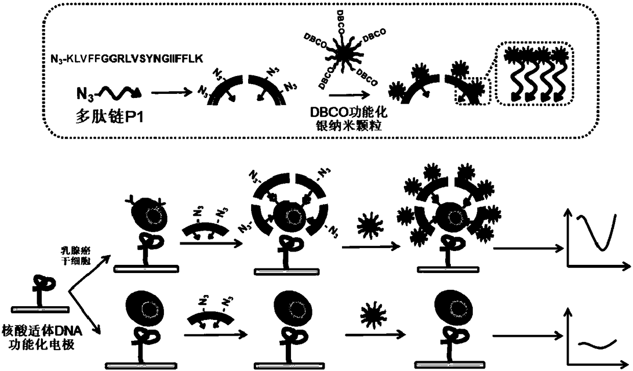
[0024] A polypeptide chain P1 modified with an azide group at the amino terminal was designed and synthesized, the sequence of which was: (NH2)Azide-KLVFFGGRLVSYNGIIFFLK(COOH). The sequence consists of three functional fragments, namely self-assembly functional fragment (KLVFF), spacer functional fragment (GG) and recognition functional fragment (RLVSYNGIIFFLK); the chemical synthesis of polypeptide chain P1 is completed by a professional peptide synthesis company.
[0025] Polypeptide chain P1 forms a polypeptide fiber by means of self-assembly functional fragments, and the azide group and recognition functional fragments are respectively located at both ends of the polypeptide fiber. The specific steps are as follows: take the stock solution of the polypeptide chain P1 in a microtube, dilute it with a certain solution, and react to make the P1 self-assemble to form a fibrous structure. The concentration of the po...
Embodiment 2
[0026] Example 2 Preparation of aptamer DNA functionalized electrode
[0027] In order to realize the capture of breast cancer stem cells, the nucleic acid aptamer DNA chain sequence should contain GGTGGTGGTGGTTGTGGTGGTGGTGG. Secondly, in order to realize the effective immobilization of the aptamer DNA chain on the electrode interface and the maintenance of capture activity, there should be multiple consecutive adenine bases at the 3' end of the sequence and a sulfhydryl group on the 6 carbon atoms (denoted as: C 6 -SH) modification. The chemical synthesis of nucleic acid aptamer DNA chain is completed by a professional DNA synthesis company.
[0028] Pretreatment of the gold electrode: First, the gold electrode is polished to a mirror surface with alumina powders with a particle size of 1 μm, 0.3 μm and 0.05 μm on the silk; then the gold electrode is placed in ethanol and ultrapure water for 3 minutes to remove residual Impurities; After rinsing with double distilled water,...
Embodiment 3
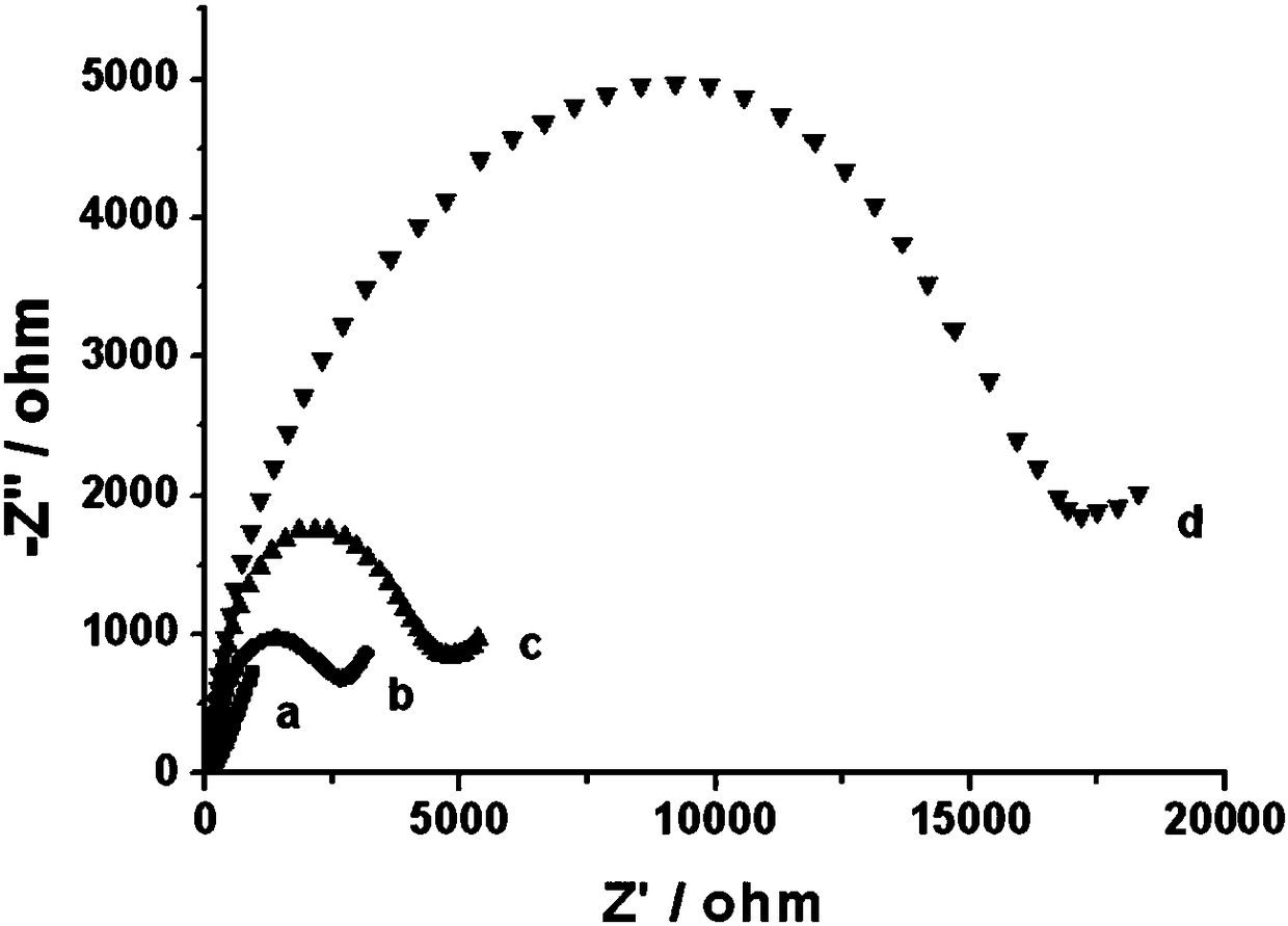
[0030] Example 3 Electrochemical sensing and detection of breast cancer stem cells
[0031] 90 μL of the sample solution containing the breast cancer stem cells to be tested interacted with the nucleic acid aptamer DNA functionalized electrode obtained in Example 2, and through the specific molecular recognition between the target cells and the nucleic acid aptamer DNA, the target breast cancer stem cells were placed on the electrode. Surface capture; the reaction time is 1 to 2 hours, and the reaction temperature is 30°C to 40°C.
[0032] The obtained electrodes capturing target breast cancer stem cells are reacted with 50 μL to 150 μL of polypeptide fibers at 30° C. to 40° C. for 1 hour to 2 hours, so as to realize the directional recognition of breast cancer stem cells by the polypeptide fibers.
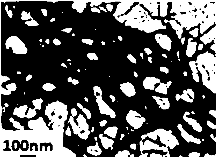
[0033]Preparation of diphenylcyclooctyne (DBCO) functionalized silver nanoparticles: 100 mL of a mixed solution containing 0.4 mM silver nitrate and 0.4 mM trisodium citrate was p...
PUM
 Abstract
Description
Claims
Application Information
 - R&D
- Intellectual Property
- Life Sciences
- Materials
- Tech Scout
- Unparalleled Data Quality
- Higher Quality Content
- 60% Fewer Hallucinations
Browse by: Latest US Patents, China's latest patents, Technical Efficacy Thesaurus, Application Domain, Technology Topic, Popular Technical Reports.
© 2025 PatSnap. All rights reserved.Legal|Privacy policy|Modern Slavery Act Transparency Statement|Sitemap|About US| Contact US: help@patsnap.com