Microarray chip preparation method, and applications of microarray chip in stem cell type brain development

A microarray chip and stem cell technology, applied in biochemical equipment and methods, tissue cell/virus culture devices, animal cells, etc., can solve the problems of restricting tissue growth and difficult removal of cell debris, so as to facilitate development and ensure Signal transmission, the effect of ensuring nutrient supply

Inactive Publication Date: 2018-06-05
DALIAN INST OF CHEM PHYSICS CHINESE ACAD OF SCI
View PDF5 Cites 7 Cited by
  • Summary
  • Abstract
  • Description
  • Claims
  • Application Information

AI Technical Summary

Problems solved by technology

At present, microfluidic chips are used to form embryoid body structures that are mostly small pits. To induce differentiation, the tissue needs to be transferred to a new low-adhesion culture dish. The main reason is that the small pit structure limits the growth of the tissue and the cell debris is difficult to remove.

Method used


Image

  • Microarray chip preparation method, and applications of microarray chip in stem cell type brain development
  • Microarray chip preparation method, and applications of microarray chip in stem cell type brain development
  • Microarray chip preparation method, and applications of microarray chip in stem cell type brain development

Examples


Embodiment 1

[0045] Fabrication of PDMS Polymer Chip with Arrayed Micropillar Structure

[0046] Use photolithography technology to make SU-8 polymer template, pour PDMS ((10~14):1 mixture) on SU-8 template, vacuum defoaming, heat in 80 degree oven for 40~60 minutes, after cooling , peel the cured PDMS off the template. Dip the unstructured PDMS block into the PDMS (20:1) on the glass slide, then seal it around the structured PDMS chip, surround the array micro-column structure to form a limited open culture pool, and place it in an 80-degree oven Heat for 60 minutes.

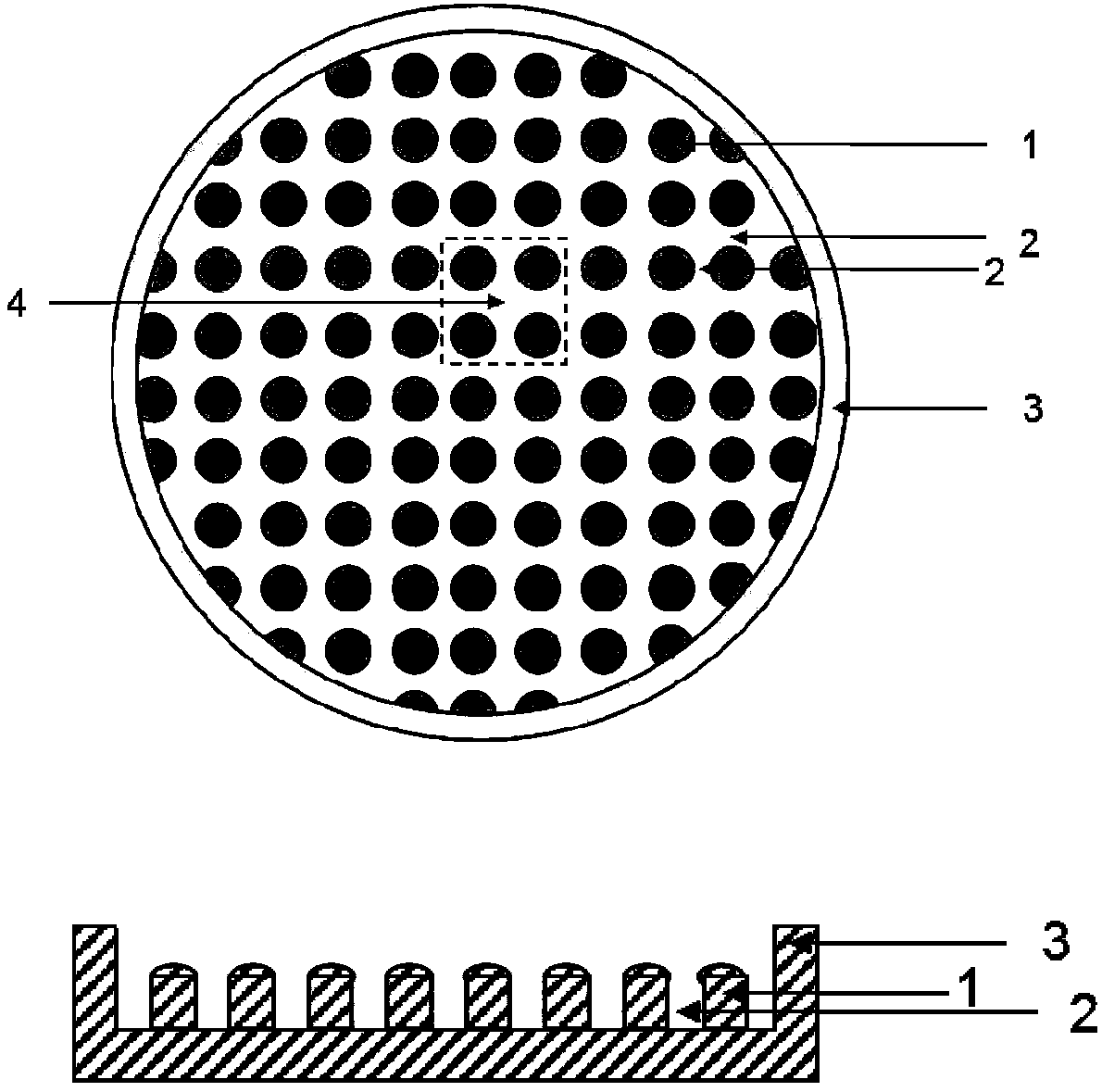
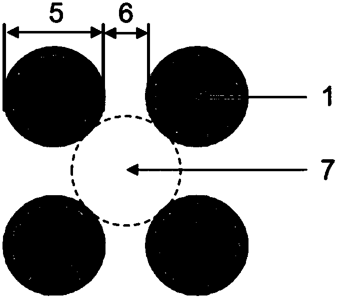
[0047] Chip structure such as figure 1 As shown, 1 is the microcolumn structure, 2 is the distance between the microcolumns, 3 is the peripheral dam structure of the chip, which is used to limit the cells and the culture medium, and 4 is the brain tissue formation area surrounded by four microcolumn structures. chip structure amplification figure 2 , 5 is the diameter of the microcolumn structure, with a length of 800 ...

Embodiment 2

[0049] High-throughput array micropillar structure chip for human induced pluripotent stem cell brain-like microtissue formation

[0050] The chip preparation process was as in Example 1. The steps of embryoid body formation and in situ brain microtissue formation are as follows:

[0051] (1) Sterilization of the chip: treat the above-mentioned microarray chip with oxygen plasma for 1-3 minutes, add deionized water; sterilize by autoclaving at 120-125°C for 20-30 minutes; place the chip in a sterile petri dish 4 Store at ℃ or use after cooling down to room temperature.

[0052] (2) Chip low-adhesion treatment: use low-adhesion reagent to treat for 4-24 hours, remove reagent and wash with PBS for 5-10 times;

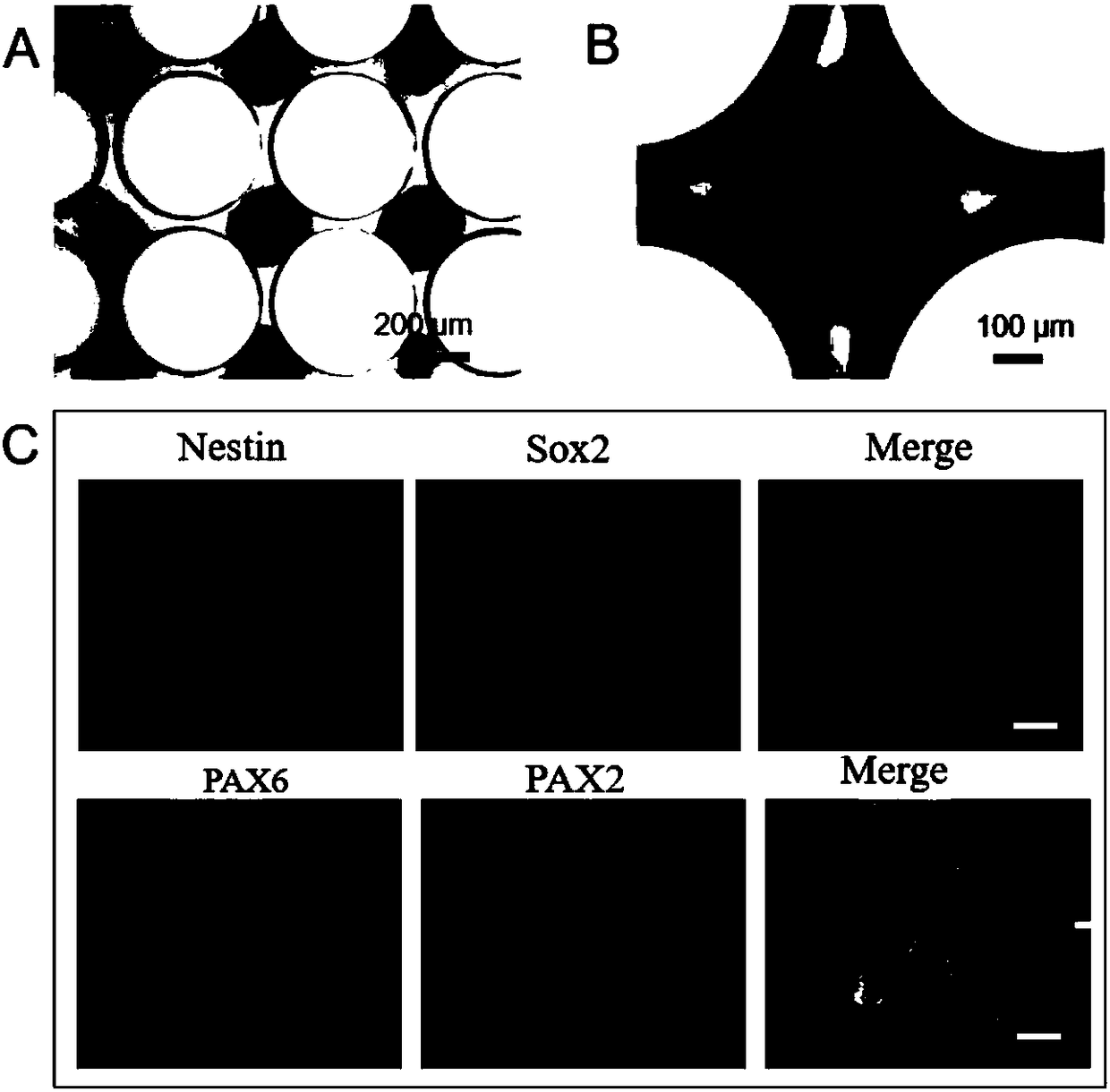
[0053] (3) Embryooid body formation: take out the chip, add mTeSR1 medium, and pipette continuously to remove the air bubbles between the small columns, so that the space between the microcolumns is filled with the medium; human induced pluripotent stem cells (hiPSCs) w...


PUM

PropertyMeasurementUnit
depthaaaaaaaaaa

Abstract

The invention provides a microarray chip preparation method, and applications of the microarray chip in stem cell type brain development, wherein the limited space enclosed by an array microcolumn structure on a chip forms a high-throughput human stem cell 3D brain tissue. With the method of the present invention, the brain-like tissues with the uniform size can be formed in the high-throughput manner, the whole growth and development process of the brain microstructures can be dynamically observed in situ, the brain tissue can be further subjected to slicing staining so as to evaluate the change of the size of the microscopic cells, and the method can simplify the experiment operation steps, dynamically observe the myocardium development in the high-throughput and in-situ manner, does notrequire special instruments and reagents, and can be integrated with other technologies; and the applications are mainly in the fields of human brain development, brain neurological disease modeling,nervous system drug and toxicological assessment, and the like.

Description

technical field [0001] The invention belongs to the field of biological technology, and in particular relates to a preparation method of a microarray chip and its application in stem cell brain-like development. . Background technique [0002] The brain is an important part of the human central nervous system and one of the important organs of the human body. Various brain diseases have seriously affected human health and quality of life. The physiological complexity of brain tissue has outstripped the capabilities of existing approaches. To study the structure and developmental characteristics of the human brain and build disease models requires new technical means. Human induced pluripotent stem cells and embryonic stem cell-derived brain tissue formation provide an important cell source for the developmental study of brain tissue and the study of brain disease mechanisms. The traditional brain tissue formation method mainly uses the three-dimensional (3D) hanging drop ...

Claims


Application Information

Patent Timeline
Patent Type & Authority Applications(China)
IPC IPC(8): C12M3/00C12N5/079C12N5/0793
CPCC12M23/02C12M23/20C12N5/0618C12N5/0619C12N2513/00
Inventor 秦建华王丽朱玉娟陶婷婷
Owner DALIAN INST OF CHEM PHYSICS CHINESE ACAD OF SCI
Why Eureka
Try Eureka