Method for evaluating lung injury caused by nanoparticles based on organ-on-a-chip technology

An organ-on-a-chip and nanoparticle technology is applied in the field of nanoparticle lung injury assessment based on organ-on-a-chip technology, achieving the effect of complete functions and simple operation.

Active Publication Date: 2018-06-05
DALIAN INST OF CHEM PHYSICS CHINESE ACAD OF SCI
View PDF4 Cites 5 Cited by
  • Summary
  • Abstract
  • Description
  • Claims
  • Application Information

AI Technical Summary

Problems solved by technology

However, it is still a blank field in lung injury research to apply it to the toxicity evaluation of nanoparticle lung i

Method used


Image

  • Method for evaluating lung injury caused by nanoparticles based on organ-on-a-chip technology
  • Method for evaluating lung injury caused by nanoparticles based on organ-on-a-chip technology
  • Method for evaluating lung injury caused by nanoparticles based on organ-on-a-chip technology

Examples


Embodiment 1

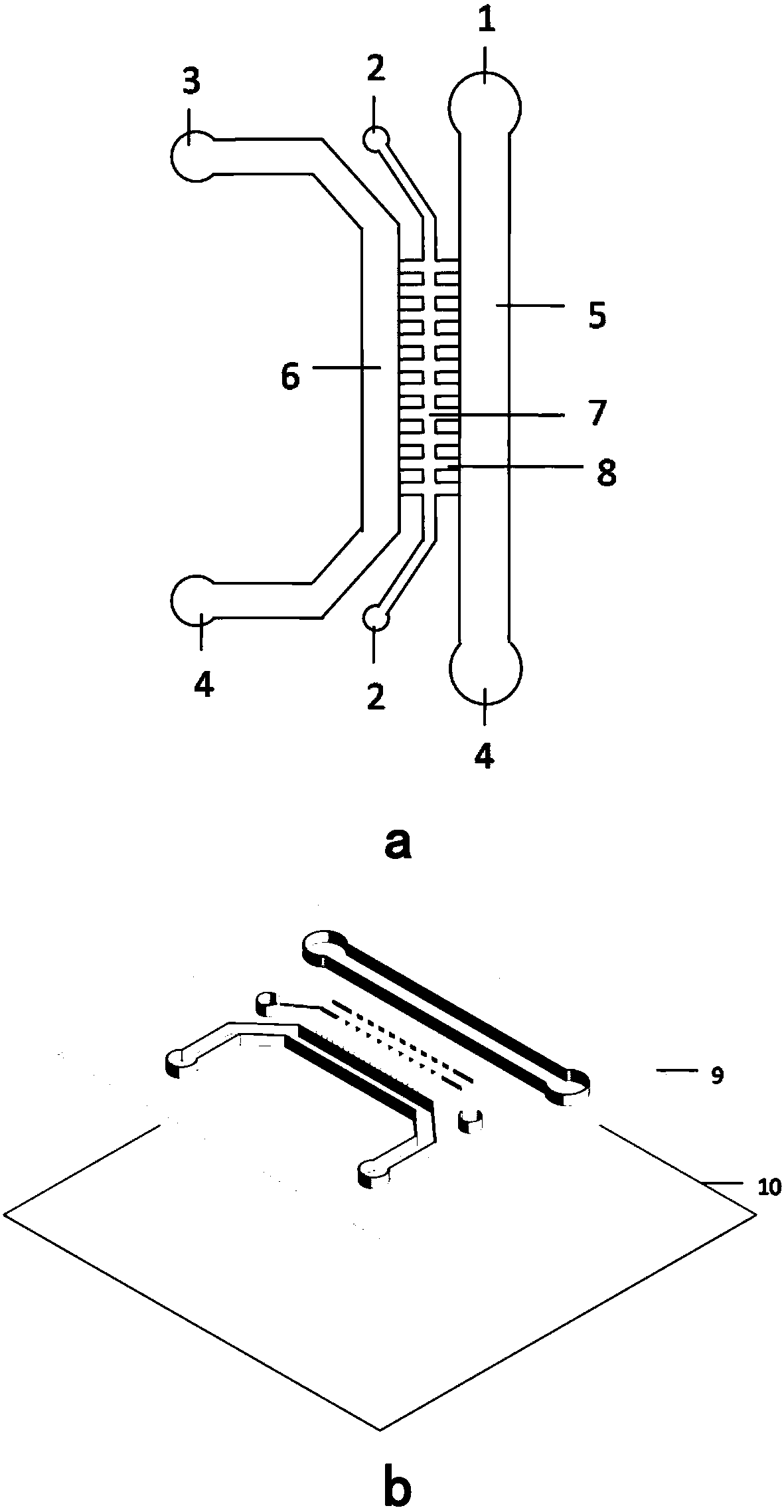
[0035] Using the microfluidic chip designed and produced by the laboratory, the configuration is shown in figure 1 . The microfluidic chip is formed by bonding and sealing the overall PDMS structure 9 on the chip upper layer and the PDMS lower layer structure 10 on the chip. Liquid pool 4, vascular endothelial cell culture room 5, alveolar epithelial cell culture room 6, stroma room 7;

[0036] The two ends of the matrix chamber 7 are extracellular matrix inlet pools, the middle part is in the shape of "Feng", and the middle horizontal structure is symmetrically arranged with 7 to 10 fence structures 8. The matrix chamber connects with the vascular endothelial cell culture chamber 5 through the fence structures on both sides. Connect with alveolar epithelial cell culture room 6;

[0037] The vascular endothelial cell culture room 5 is connected to the vascular endothelial cell inlet pool 1 on the upper side and the waste liquid pool 4 on the lower side; the alveolar epitheli...

Embodiment 2

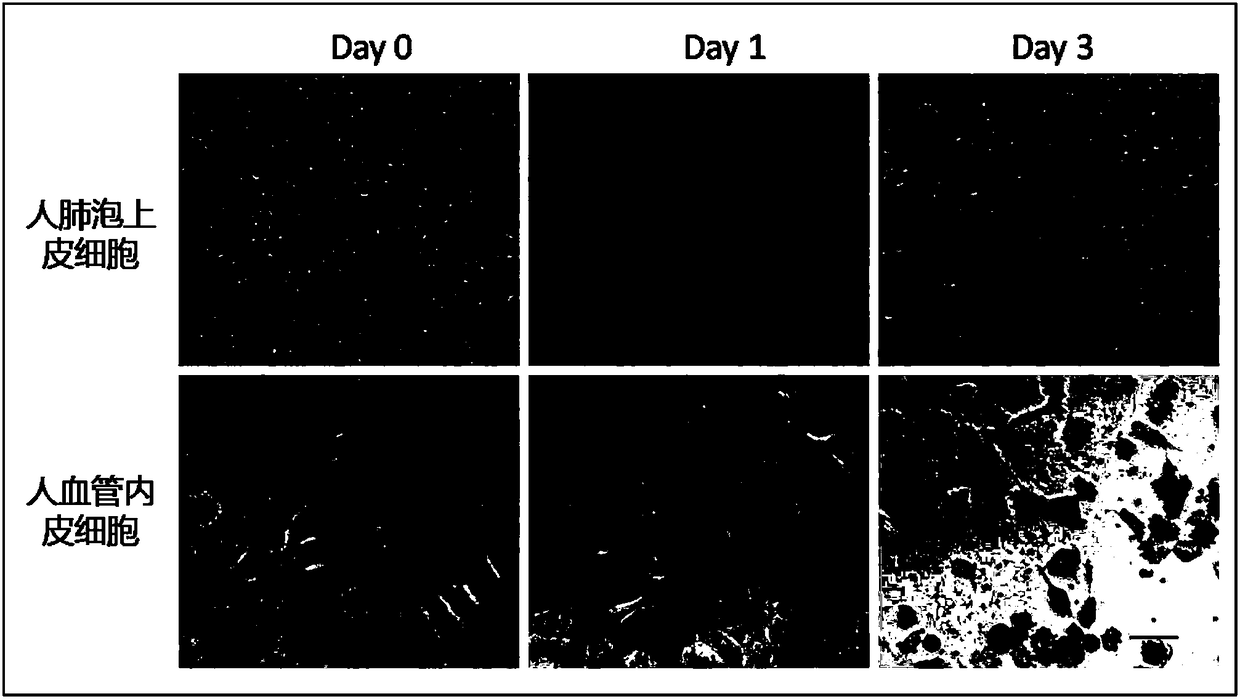
[0040] A nanoparticle-based lung injury assessment method based on organ-on-a-chip technology. The specific process is as follows:

[0041] Prepare matrigel working solution, add it to the matrix inlet pool with a pipette, 1 μl per well; add 1ml PBS buffer solution to the culture dish, put the culture dish with the fixed chip into the incubator and incubate the gel for 30 minutes to promote the formation of matrigel from the sticky The thick liquid turns into a jelly-like gel. After the gel process is over, add the fresh culture medium of the two kinds of cells from the inlet pool of vascular endothelial cells and the inlet pool of alveolar epithelial cells respectively; make the suspension of vascular endothelial cells, take 10 μl The liquid was added to the inlet pool of vascular endothelial cells, and 10 μl of cell culture liquid was quickly sucked from the waste pool to promote the cells to enter the cell culture chamber quickly and evenly under the action of gravity flow....

Embodiment 3

[0043] Fabricate a microfluidic chip, consisting of upper and lower layers of PDMS, including vascular endothelial cell inlet pool, extracellular matrix inlet pool, alveolar epithelial cell inlet pool, waste liquid pool, vascular endothelial cell culture chamber, alveolar epithelial cell culture chamber and matrix chamber and other structures. The height of the vascular endothelial cell culture chamber and the alveolar epithelial cell culture chamber is 200-1000 μm, and the height of the stromal inlet pool and the stromal chamber is 200 μm. Add 1 μl of matrigel working solution into the matrix inlet pool, and incubate at 30°C for 30 minutes. Add 20 μl of vascular endothelial cell suspension into the inlet pool of vascular endothelial cells, erect the chip at 90 degrees, and place it in a 37° C. incubator for 6 hours. The same method was used to inoculate lung epithelial cells in the lung epithelial cell culture room and stand upright for 1 hour, then put the chip flat and mov...


PUM


Abstract

The invention provides a method for evaluating lung injury caused by nanoparticles based on organ-on-a-chip technology. A chip is mainly prepared by bonding and sealing an upper PDMS layer and a lowerPDMS layer and comprises a matrix inlet pool, two cell inlet pools, a waste liquid pool, a cell culture chamber and a matrix chamber. The chip is vertically inoculated with vascular endothelial cellsand alveolar epithelial cells separately, and culture is carried out for a certain period of time so as to form a lung air-blood barrier. Nanometers are added into an alveolar epithelial cell culturechamber, and monocytes are added into a vascular endothelial cell culture chamber in the manner of perfusion to simulate monocytes in the circulating blood of people, so an in-vitro nanoparticle lunginjury evaluation model is established. The nanoparticle lung injury evaluation model based on the chip can simulate the injury of the nanoparticles to the lung air-blood barrier, monitors destroy tothe integrity of the barrier in real time, realizes observation of the responses of two types of cells of the air-blood barrier, and traces the motion of the monocytes in real time.

Description

technical field [0001] The invention relates to the technical field of applying organ chip technology to the in vitro evaluation of nanoparticle lung injury, in particular to a nanoparticle lung injury evaluation method based on organ chip technology. Background technique [0002] With the acceleration of economic development and urbanization, regional air pollution characterized by atmospheric particulate matter and ozone has become the most important environmental factor affecting the health of Chinese residents at this stage. Atmospheric particulate matter is a general term for various solid and liquid granular substances existing in the atmosphere. The harm to human health is related to its particle size. The smaller the particle size, the greater the harm. The lung is the main organ of the respiratory system. As a part directly in contact with the atmospheric environment, the lung-air-blood barrier is the lung's first line of defense against fine particle pollution. At...

Claims


Application Information

Patent Timeline
IPC IPC(8): C12M3/00C12Q1/02
CPCC12M21/08C12M23/16C12M35/08C12N2503/04G01N33/5026G01N33/5029G01N33/5088
Inventor 秦建华张敏
Owner DALIAN INST OF CHEM PHYSICS CHINESE ACAD OF SCI
Why Eureka
Try Eureka