Detection kit for heparin binding protein and preparation method thereof

A technology of heparin-binding protein and detection kit, which is applied in the detection field of heparin-binding protein, can solve the problems of detection reagent sensitivity, specificity, and unsatisfactory linear range, and achieves the effects of wide measurement linear range, simple operation and simple composition.

Inactive Publication Date: 2018-06-12
SUZHOU KANGHESHUN MEDICAL TECH
View PDF4 Cites 11 Cited by
  • Summary
  • Abstract
  • Description
  • Claims
  • Application Information

AI Technical Summary

Problems solved by technology

Many problems make the detection reagent obtained by this invention unsatisfactory in terms of sensitivity, specificity, linear range, etc., and the accuracy of the reagent cannot be well correlated with existing marketed reagents

Method used


Image

  • Detection kit for heparin binding protein and preparation method thereof
  • Detection kit for heparin binding protein and preparation method thereof
  • Detection kit for heparin binding protein and preparation method thereof

Examples


Embodiment 1

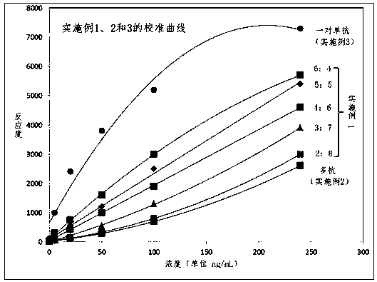
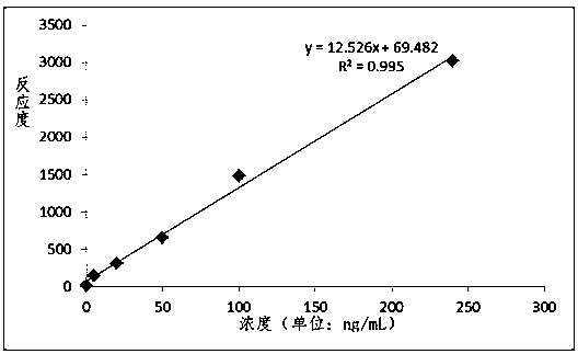
[0045] 1) Preparation of latex reagent (333nm, reaction solution R2) labeled with combination of monoclonal antibody and polyclonal antibody

[0046] Polystyrene latex particles with carboxyl groups on the surface (classification number P0323, manufacturer JSR Life Sciences Corporation) with a particle size of 333 nm were labeled by the intermediate ester method. The surface carboxylated microparticles with a particle size of 333 nm were diluted to 1% in MES buffer solution and stirred at room temperature. Throw in EDC and Sulfo-NHS solid powder, centrifuge at 10000RPM for 20 minutes after stirring for 2 hours. The latex was washed twice with MES buffer solution, resuspended and divided into two parts. One part was added with HBP monoclonal antibody, stirred for 2 hours, and the other part was added with HBP polyclonal antibody, stirred for 2 hours. Both latexes were then centrifuged at 10,000 RPM for 20 minutes, and the supernatant was discarded. The two sets of precipitat...

Embodiment 2

[0056] 1) Preparation of polyantibody-labeled latex reagent (333nm, reaction solution R2)

[0057] Polystyrene latex particles with carboxyl groups on the surface (classification number P0323, manufacturer JSR Life Sciences Corporation) with a particle size of 333 nm were labeled by the intermediate ester method. The surface carboxylated microparticles with a particle size of 333 nm were diluted to 1% in MES buffer solution and stirred at room temperature. Throw in EDC and Sulfo-NHS solid powder, centrifuge at 10000RPM for 20 minutes after stirring for 2 hours. The latex was then centrifuged at 10,000 RPM for 10 minutes, and the supernatant was discarded. The precipitated latex was resuspended in the stock solution and ultrasonically dispersed, stirred for 1 hour before use. In this example, reaction solution R2 includes latex particles, buffer solution, salt, stabilizer, suspending agent and preservative, wherein the concentration of latex particles labeled with HBP antibod...

Embodiment 3

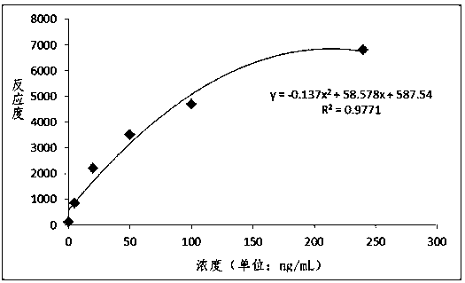
[0060] 1) Preparation of latex reagent (333nm, reaction solution R2) of a pair of monoclonal antibodies

[0061]Polystyrene latex particles with carboxyl groups on the surface (classification number P0323, manufacturer JSR Life Sciences Corporation) with a particle size of 333 nm were labeled by the intermediate ester method. The surface carboxylated microparticles with a particle size of 333 nm were diluted to 1% in MES buffer solution and stirred at room temperature. Throw in EDC and Sulfo-NHS solid powder, centrifuge at 10000RPM for 20 minutes after stirring for 2 hours. The latex was washed twice with MES buffer solution, resuspended and divided into two parts. Each of the HBP monoclonal antibodies was added, and the reaction was stirred for 2 hours. Both latexes were then centrifuged at 10,000 RPM for 20 minutes, and the supernatant was discarded. Both precipitated latexes were resuspended and sonicated in stock solution, and stirred separately for 1 hour. In this exa...


PUM

PropertyMeasurementUnit
Diameteraaaaaaaaaa
Particle sizeaaaaaaaaaa

Abstract

The invention discloses a detection kit for heparin binding protein and a preparation method thereof. The kit is a latex-enhanced immunoturbidimetry detection reagent. The latex-enhanced immunoturbidimetric detection reagent is prepared from the following main components of: buffering solution, a surface active agent, diluted liquid R1 of salt and preservative, and reaction liquid R2 containing latex particles labeled by HBP (Heparin Binding Protein) antibodies, buffering solution, the salt, a stabilizer, a suspending agent and the preservative, HBP calibration products and quality control products. The invention also discloses a method for detecting the concentration of the heparin binding protein (HBP) in a blood sample by using the kit and utilizing the transmitting or scattering turbidimetric principle. The detection kit disclosed by the invention adopts dual reagents, is simple in operation, high in sensitivity and wide in linear range, can be widely applied to various transmitting or scattering analyzers including common biochemical analyzers and specific protein analyzers and the like.

Description

technical field [0001] The invention belongs to the field of medical immunodiagnostic reagents, in particular to the detection of heparin binding protein. Background technique [0002] Heparin Binding Protein (HBP for short) is a granular protein secreted by neutrophils, which has a bactericidal effect and is also involved in the regulation of inflammatory response and blood coagulation process. In 1984, Shafer et al. first discovered and isolated this protein, and named it CAP37 (Cationic Antimicrobial Protein, molecular weight 37k) according to its electropositive characteristics and bactericidal function. Later, azurocidin and heparin-binding protein (HBP-Heparin Binding Protein), which has a strong binding ability to heparin, were isolated from the azurophilic blue granules in polymorphonuclear leukocytes. Further studies on protein structure and gene sequencing show that these three proteins are actually the same protein, which is represented by heparin-binding protein...

Claims


Application Information

Patent Timeline
IPC IPC(8): G01N33/68G01N33/532
CPCG01N33/6893G01N33/532
Inventor 陈胜胜刘向晖王明
Owner SUZHOU KANGHESHUN MEDICAL TECH
Why Eureka
Try Eureka