Brain tissue membrane protein extraction method

An extraction method and membrane protein technology, which is applied in the field of membrane protein extraction, can solve the problems of protein denaturation, loss of protease activity and channel activity, and cannot be restored, and achieve the effect of efficient extraction and simple system formula

Inactive Publication Date: 2018-08-10
KUNMING MEDICAL UNIVERSITY
View PDF4 Cites 4 Cited by
  • Summary
  • Abstract
  • Description
  • Claims
  • Application Information

AI Technical Summary

Problems solved by technology

However, if the concentration used is high, there will be two major problems: one is that the protein is severely denatured, the structure changes and cannot be restored.
In this case, the general determination of the amount of the protein will not be a problem, but the enzymat...

Method used


Image

  • Brain tissue membrane protein extraction method
  • Brain tissue membrane protein extraction method
  • Brain tissue membrane protein extraction method

Examples


Embodiment 1

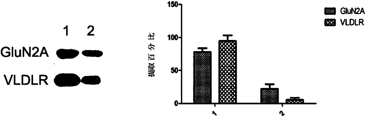
[0030] Such as figure 1 As shown, the present invention is used to extract two membrane-integrated proteins, namely GluN2A protein and VLDLR protein, and the undissolved precipitate is further lysed with a strong RIPA lysate to extract the unextracted protein of the present invention . The two parts of the protein were electrophoresed by SDS-PAGE in the same ratio, and it can be seen that more than 78% of GLuN2A and more than 95% of VLDLR were extracted by using the present invention.

Embodiment 2

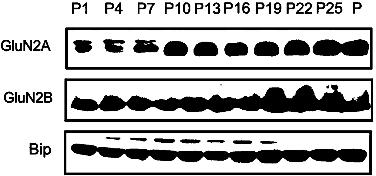
[0032] Using the present invention to extract membrane protein expression in mouse brain tissue at different stages of development and analyze:

[0033] 1. Take mice of different ages, namely mice and female mice born at 1, 4, 7, 10, 13, 16, 19, 22, and 25 days old.

[0034] 2. The mice were killed by neck dislocation, the brain tissue was quickly removed, and the cortex was separated.

[0035] 3. Weigh the isolated cortical tissue, add 1ml of solution b pre-cooled to 4°C for every 100mg of tissue, and cut the tissue into small pieces of 1 mm cube with ophthalmic scissors. Homogenize with a tissue homogenizer. Use Thermo's cocktail mix of protease inhibitors. Thermo Fisher, Protease Inhibitor Cocktail (100X), Cat. No.: 78430.

[0036] 4. Centrifuge the homogenized tissue suspension with a centrifugal force of 700g at 4°C for 10 minutes, take out the supernatant and transfer it to a new centrifuge tube, and discard the precipitate.

[0037] 5. Add 0.1 times the volume of so...

Embodiment 3

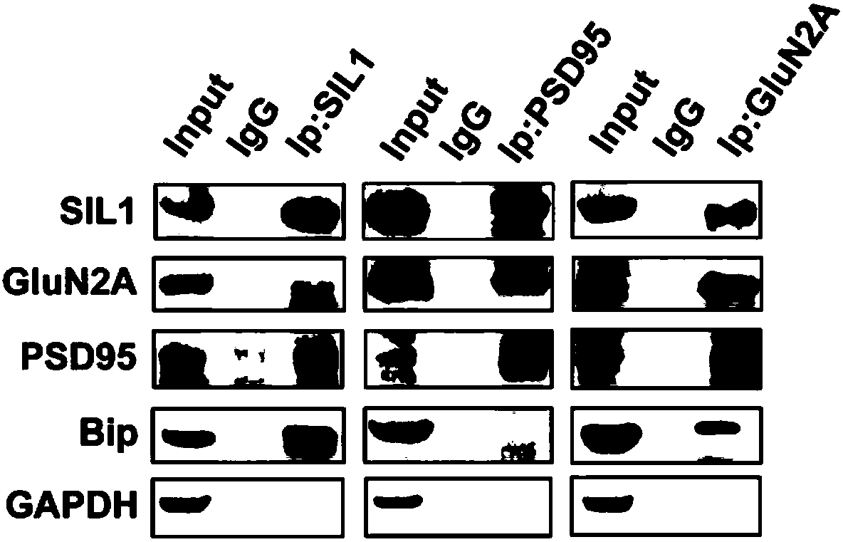
[0046] After using the present invention to extract proteins, the interaction between proteins was analyzed using co-immunoprecipitation method:

[0047] 1. Take mice of different ages, namely mice and female mice born at 1, 4, 7, 10, 13, 16, 19, 22, and 25 days old.

[0048] 2. The mice were killed by neck dislocation, the brain tissue was quickly removed, and the cortex was separated.

[0049] 3. Weigh the isolated cortical tissue, add 1ml of solution b pre-cooled to 4°C for every 100mg of tissue, and cut the tissue into small pieces of 1 mm cube with ophthalmic scissors. Homogenize with a tissue homogenizer. Use Thermo's cocktail mix of protease inhibitors. Thermo Fisher, Protease Inhibitor Cocktail (100X), Cat. No.: 78430.

[0050] 4. Centrifuge the homogenized tissue suspension with a centrifugal force of 700g at 4°C for 10 minutes, take out the supernatant and transfer it to a new centrifuge tube, and discard the precipitate.

[0051] 5. Add 0.1 times the volume of sol...


PUM


Abstract

The invention provides a brain tissue membrane protein extraction method, and relates to the technical field of biology. A used scale removal agent is very mild; in addition, the concentration is verylow; a reducing agent and a protein allosteric agent are not used; the protein structure and activity can be maintained. Membrane lipid surrounding the extracted protein is not completely lost; a tertiary structure of the protein can be effectively remained, so that the combination state of the protein with other protein can be stabilized. By using the extraction method provided by the invention,80 percent of membrane protein or more can be effectively dissolved. The formula of a used buffer solution system is simple; only conventional reagents and instrument equipment in a laboratory are needed; the result and the activity of the protein are not influenced at all by the used stain removal agent and the dosage; the extracted membrane protein can be used for biochemical tests with higherdifficulty in co-immunoprecipitation and the like and higher protein extraction requirements.

Description

technical field [0001] The invention relates to the field of biotechnology, in particular to a membrane protein extraction method. Background technique [0002] Animal cells contain a lot of protein. Proteins can be divided into two categories according to their relationship with the cell membrane, namely cytoplasmic proteins and membrane proteins. The cytoplasmic protein is a soluble protein, which exists in the cytoplasm in a free state. Membrane proteins can be further divided into three categories: peripheral proteins, integral proteins, and lipid-anchored proteins. Peripheral proteins are bound to the membrane by hydrogen bonds, etc., and lipid-anchored proteins are inserted into the membrane lipid bilayer through the lipid sequence connected on the protein. The combination of these two types of membrane proteins and the membrane is relatively loose. Integrins, on the other hand, have a hydrophobic (lipophilic) sequence and a hydrophilic sequence. It inserts on the ...

Claims


Application Information

Patent Timeline
IPC IPC(8): C07K14/47C07K1/34
CPCC07K14/47
Inventor 张筱敏徐世莲杨媛
Owner KUNMING MEDICAL UNIVERSITY
Why Eureka
Try Eureka