Preparation method of gypsum scanning electron microscope sample

A scanning electron microscope and gypsum technology, applied in the direction of measuring devices, instruments, scientific instruments, etc., can solve the problems of not being able to prepare gypsum samples well, not being able to obtain high-quality microcrystalline morphology photos, and not being able to obtain sizes, etc., to achieve Good electrical conductivity, uniform thickness, fast sample preparation

Pending Publication Date: 2018-10-19
SHANDONG JIANZHU UNIV
View PDF8 Cites 0 Cited by
  • Summary
  • Abstract
  • Description
  • Claims
  • Application Information

AI Technical Summary

Problems solved by technology

However, because gypsum is a brittle material, neither of the above two methods can obtain gypsum samples with small and regular size, smooth surface, and undamaged microcrystalline morphology; Obtain high-quality, clear images of crystallite topography
[0004] In summary, the existing methods for preparing gypsum SE

Method used


Image

  • Preparation method of gypsum scanning electron microscope sample
  • Preparation method of gypsum scanning electron microscope sample
  • Preparation method of gypsum scanning electron microscope sample

Examples


Embodiment 1

[0053] A preparation method for a gypsum scanning electron microscope sample, comprising the steps of:

[0054] 1) Mix water and building gypsum powder of no less than 200 meshes, the ratio of mixture to water is 100:55, and stir quickly to form a uniform slurry. The stirring time is 1min. The slurry is viscous but can flow.

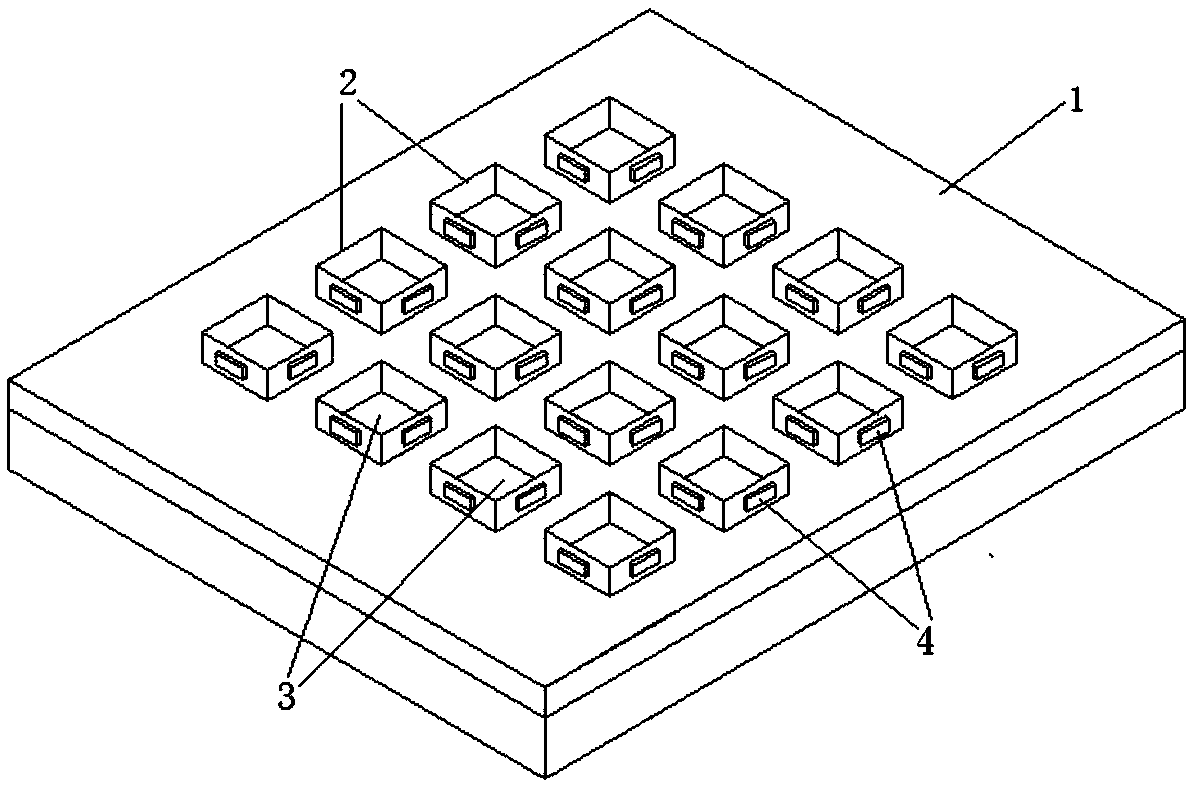
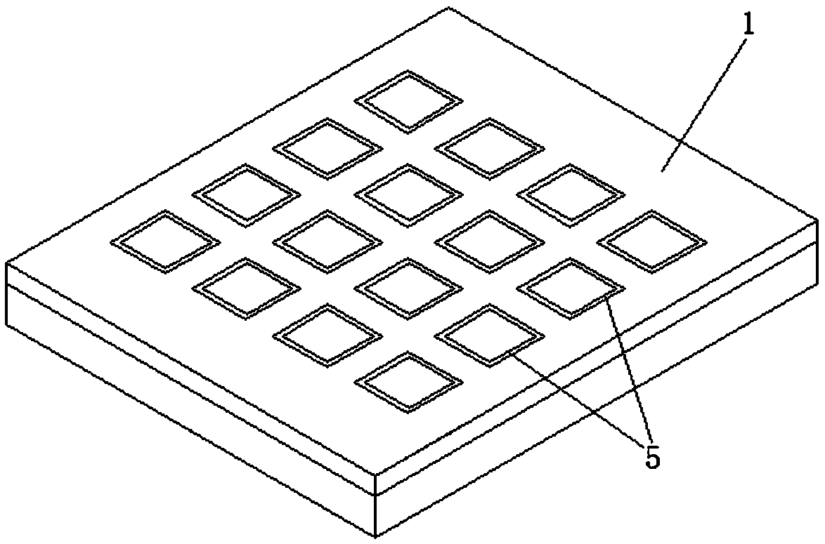
[0055] 2) Pour the slurry in step 1) into the cavity of the mold used to prepare the gypsum scanning electron microscope sample, scrape it flat, dry it at 25°C for 2 hours, and demould after molding to obtain a flaky gypsum sample; Mold such as figure 1 with 2 As shown, it includes a base plate 1, a baffle plate 2, a handle 4, and a draw slot 5. There are 16 draw slots distributed on the base plate 1, and the width of the baffle plate 2 matches the width of the draw slot 5, so that the baffle plate 2 Can be tightly engaged in the card slot 5, the baffle plate 2 is engaged in the card slot 5 to form the inner cavity 3, the size of the inner cavity 3 is ...

Embodiment 2

[0061] A preparation method for a gypsum scanning electron microscope sample, comprising the steps of:

[0062] 1) Mix water with not less than 200 meshes of building gypsum and fly ash powder, the ratio of the mixture to water is 100:53, stir quickly to form a uniform slurry, the stirring time is 2min, the slurry is viscous but can flow ;

[0063] 2) Pour the slurry in step 1) into the cavity of the mold used to prepare the gypsum SEM sample, scrape it flat, dry it at 25°C for 3 hours, and demould after molding to obtain a thin slice of 5mm×5mm×1mm Shape gypsum sample; The mold that adopts is with embodiment 1.

[0064] 3) Curing the gypsum sample in step 2) in a curing box, the curing temperature is 22°C, the curing humidity is 95%, and the curing time is 28 days;

[0065] 4) Put the gypsum sample cured in step 3) into an oven for drying, the drying temperature is 45°C, and the drying time is 24 hours;

[0066] 5) Paste the conductive tape on the side of the cuboid alumin...

Embodiment 3

[0069] A preparation method for a gypsum scanning electron microscope sample, comprising the steps of:

[0070] 1) Mix water with not less than 200 meshes of building gypsum, cement, and silica fume powder. The ratio of the powder mixture to water is 100:52. Stir quickly to form a uniform slurry. The stirring time is 2 minutes, and the slurry is viscous. but can flow;

[0071] 2) Pour the slurry in step 1) into the cavity of the mold used to prepare the gypsum scanning electron microscope sample, scrape it flat, dry it at 25° C. for 2 hours, and demould after molding to obtain a thin sheet of gypsum sample; Mold is with embodiment 1.

[0072] 3) Curing the gypsum sample in step 2) in a curing box, the curing temperature is 22°C, the curing humidity is 95%, and the curing time is 28 days;

[0073] 4) Put the gypsum sample cured in step 3) into an oven for drying, the drying temperature is 45°C, and the drying time is 24 hours;

[0074] 5) Paste the conductive tape on the sid...


PUM

PropertyMeasurementUnit
Finenessaaaaaaaaaa

Abstract

Belonging to the technical field of preparation of scanning electron microscope samples, the invention relates to a preparation method of a gypsum scanning electron microscope sample. The preparationmethod comprises the steps of: 1) blending water and a gypsum powder raw material into a homogeneous slurry; 2) pouring the well blended slurry of step 1) into a mold, scraping the slurry flat, makingthe slurry surface flush with the upper mouth margin of a die, and then conducting drying, molding, demoulding, curing and drying to obtain a gypsum test block; 3) pasting a conductive adhesive tapeto a sample table, and then pasting the gypsum test block of step 2) on the conductive adhesive tape; and 4) using inert gas to purge the upper surface of the sample table pasted with the gypsum testblock of step 3), then conducting spray coating of a conductive metal. The gypsum sample prepared by the method provided by the invention has small size, is regular, thin and uniform, and has a smoothsurface, and the microcrystalline morphology is not destroyed, and the microcrystalline morphology photograph obtained by the gypsum sample under a scanning electron microscope is free of false and fuzzy phenomena. At the same time, the preparation method provided by the invention has the advantages of simple operation, rapid sample preparation and high efficiency.

Description

technical field [0001] The invention belongs to the technical field of preparation of scanning electron microscope samples, in particular to a preparation method of gypsum scanning electron microscope samples. Background technique [0002] The observation of the microcrystalline morphology of inorganic gelling material products often requires the help of a microscope that can magnify more than a thousand times, and scanning electron microscopy is one of the most commonly used equipment. It mainly uses secondary electron signal imaging to observe the surface morphology of the sample, that is, to scan the sample with a very narrow electron beam, and to produce various effects through the interaction between the electron beam and the sample. Based on this, SEM has strict requirements on the conductivity of the sample. For gypsum products, which are non-conductive and porous inorganic gelling materials, samples with good conductivity must be prepared in order to observe their m...

Claims


Application Information

Patent Timeline
IPC IPC(8): G01N23/2202
CPCG01N23/2202
Inventor 井敏朱孟晗宋宪涛徐丽娜岳雪涛
Owner SHANDONG JIANZHU UNIV
Why Eureka
Try Eureka