Immumofluorescence method kit used for detecting placental growth factor
A placental growth factor and immunofluorescence technology, which is applied in the field of immunofluorescence kits for placental growth factor detection, can solve the problems of the need for replacement of electrodes, insufficient sensitivity, high background signal, etc., so as to eliminate the interference of non-specific fluorescence and improve the High analytical sensitivity and high fluorescence intensity
- Summary
- Abstract
- Description
- Claims
- Application Information
AI Technical Summary
Problems solved by technology
Method used
Image

Examples
Embodiment 1
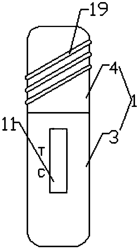
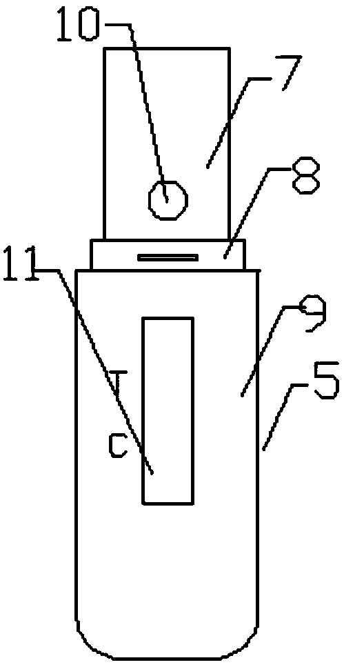
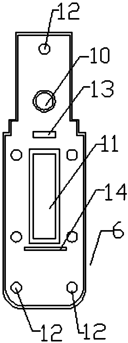
[0030] Such as Figure 1-8As shown, an immunofluorescence kit for detection of placental growth factor comprises a kit box body 1 and a test strip 2 arranged in the kit box body, the kit box body includes a housing 3, a protective cover 4, The housing 3 and the protective sleeve 4 are connected by clamping. The housing 3 includes an upper housing 5 and a lower housing 6. The upper housing 5 includes an upper housing upper part 7, an upper housing middle part 8, and an upper housing whose width increases successively. The lower part 9, the upper part of the upper casing is provided with a sample injection hole 10, the sample injection hole 10 is a circular structure, the lower part of the upper casing 9 is provided with a detection window 11, and the detection window 11 is a rectangular structure, and the inner surface of the upper casing 5 is provided with There are a first card body 12, a first card strip 13, and a second card strip 14, the first card body 12 is a cylindrical...
Embodiment 2
[0047] This embodiment is basically the same as Embodiment 1, the difference is:
[0048] A preparation method for immunofluorescence test strips for placental growth factor detection, comprising the steps of:
[0049] (1) Preparation of placental growth factor monoclonal antibody conjugates labeled with europium fluorescent nanospheres: dissolve europium fluorescent nanospheres in CBS buffer (50 mM, pH=9.6), wash 3 times by centrifugation, and the centrifugation speed is 18000rpm for 3 minutes, finally resuspend in CBS buffer (50mM, pH=9.6), add aldylated dextran, mix well, react in dark at room temperature for 5 hours, after the reaction is completed, use the above centrifugation method Wash and then resuspend in CBS buffer (50mM, pH=9.6) to obtain formylated europium fluorescent nanospheres, which are stored at 4°C for later use; placenta growth factor monoclonal antibody conjugate labeled with europium fluorescent nanospheres Preparation of placental growth factor monoclo...
PUM
| Property | Measurement | Unit |
|---|---|---|
| Diameter | aaaaa | aaaaa |
Abstract
Description
Claims
Application Information
 - Generate Ideas
- Intellectual Property
- Life Sciences
- Materials
- Tech Scout
- Unparalleled Data Quality
- Higher Quality Content
- 60% Fewer Hallucinations
Browse by: Latest US Patents, China's latest patents, Technical Efficacy Thesaurus, Application Domain, Technology Topic, Popular Technical Reports.
© 2025 PatSnap. All rights reserved.Legal|Privacy policy|Modern Slavery Act Transparency Statement|Sitemap|About US| Contact US: help@patsnap.com