Preparation method of octahedral 3D printed PLGA/hydroxyapatite cervical interbody fusion cage, product and application thereof

A technology of hydroxyapatite and intervertebral fusion, which is applied in the field of medical materials, can solve the problems of patients with foreign bodies, large opening trauma, cumbersome operation, etc., and achieve the effects of convenient implantation, accelerated fusion, and increased contact area

Inactive Publication Date: 2018-12-04
SHANGHAI NAT ENG RES CENT FORNANOTECH
View PDF10 Cites 5 Cited by
  • Summary
  • Abstract
  • Description
  • Claims
  • Application Information

AI Technical Summary

Problems solved by technology

The intervertebral fusion device on the market has a large opening trauma during operation, and the opera

Method used


Image

  • Preparation method of octahedral 3D printed PLGA/hydroxyapatite cervical interbody fusion cage, product and application thereof
  • Preparation method of octahedral 3D printed PLGA/hydroxyapatite cervical interbody fusion cage, product and application thereof
  • Preparation method of octahedral 3D printed PLGA/hydroxyapatite cervical interbody fusion cage, product and application thereof

Examples


Embodiment 1

[0046]A preparation method of an octahedral 3D printed PLGA / hydroxyapatite cervical intervertebral fusion device, using 3D printing technology, and polylactic acid-polyglycolic acid (PLGA) copolymer as the biomaterial of the cervical intervertebral fusion device Stent main body 1, preparing biodegradable nano-hydroxyapatite composite fusion cage stent by fused deposition forming 3D printing, includes the following steps:

[0047] (1) Preparation of PLGA / hydroxyapatite composite:

[0048] Cut the PLGA with a molecular weight of 200,000 into small pieces, put it into the printing chamber of the 3D plotter preheated at 140°C, the stacking height between the 3D layers is 0.5 mm, and 3D print to make the neck fusion device;

[0049] (2) Preparation of cervical intervertebral fusion cage model:

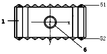
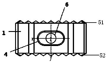
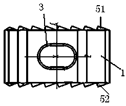
[0050] see Figure 1-Figure 6 , use CAD software to edit the finite element model based on the real anatomical model of the finite element human cervical spine, save it as a stl standard ...

Embodiment 2

[0058] Similar to Example 1, except that the nano-hydroxyapatite powder in PLGA / hydroxyapatite is 10% by mass.

[0059] First, cut PLGA (200,000 molecular weight) into small pieces and fill them into the cavity of a Haake rheometer preheated to 140 degrees Celsius, add 10% by mass fraction of nano-hydroxyapatite powder in stages, and mix thoroughly for about After 1 hour, after cooling to 70°, disassemble the cavity and take out the material. Cut the mixed material into small pieces with scissors and stuff them into the printing chamber of the 3D plotter, set the temperature to 140 degrees Celsius for 30 minutes, set the stacking height between layers to 0.5 mm, and print to make the neck fuser.

[0060] For cervical fusion cage data model, see Figure 1-Figure 6 , this embodiment mainly includes that the main body 1 has a similar octagonal cylinder structure, and the asymmetrical octagonal cylinder shape is beneficial to the surgical operation of cervical fusion cage implant...

Embodiment 3

[0067] Similar to Example 1, except that the nano-hydroxyapatite powder in PLGA / hydroxyapatite is 20% by mass.

[0068] First, cut PLGA (200,000 molecular weight) into small pieces and fill them into the cavity of a Haake rheometer preheated to 140 degrees Celsius, add 20% by mass fraction of nano-hydroxyapatite powder in stages, and mix thoroughly for about After 1 hour, after cooling to 70°, disassemble the cavity and take out the material. Cut the mixed material into small pieces with scissors and stuff them into the printing chamber of the 3D plotter, set the temperature to 140 degrees Celsius for 30 minutes, set the stacking height between layers to 0.5 mm, and print to make the neck fuser.

[0069] For cervical fusion cage data model, see Figure 1-Figure 6 , this embodiment mainly includes that the main body 1 has a similar octagonal cylinder structure, and the asymmetrical octagonal cylinder shape is beneficial to the surgical operation of cervical fusion cage implant...


PUM


Abstract

The invention relates to a preparation method of an octahedral 3D printed PLGA/hydroxyapatite cervical interbody fusion cage, a product and an application thereof. A solid part takes polylactic acid-polyglycolic acid (PLGA) copolymer as a main component to be a main component of a biological scaffold, and the nano-sized hydroxyapatite particles are used as a filling inorganic material, The biodegradable nano-hydroxyapatite composite fusion cage is prepared by fused deposition 3D printing, the method comprises preparation of a PLGA/hydroxyapatite composite material, preparation of a cage modeland application of 3D plotter bioprinter for fused deposition to prepare cervical fusion cage. The cervical interbody fusion cage of the invention is convenient to implant, accelerates the fusion of the device and the cervical vertebra, and is not easy to slide out of an implantation site and has high biocompatibility, a bone graft aperture is filled with a bone graft, the fusion of tissue and materials can be accelerated, the cavity is filled with new bone cells, and the fusion effect is enhanced.

Description

technical field [0001] The invention relates to a preparation method, product and application of an octahedral 3D printed PLGA / hydroxyapatite cervical intervertebral fusion device, belonging to the field of medical material technology applications. technical background [0002] Cervical spondylosis is the degenerative change of cervical intervertebral disc tissue and its secondary and involves cervical nerve roots, spinal cord, vertebral artery and foot shaft nerves, which seriously affects the life and work of patients. For patients with cervical spondylosis who fail conservative treatment, surgical treatment may be implemented if necessary according to the actual condition of the disease. Currently, anterior intervertebral fusion and fixation are the main means of treating cervical spondylosis such as traumatic disc herniation, osteophyte hyperplasia at the posterior border of the vertebral body, and limited ossification of the posterior longitudinal ligament. Cervical in...

Claims


Application Information

Patent Timeline
IPC IPC(8): A61L27/12A61L27/18A61L27/50B33Y10/00B33Y80/00A61F2/44
CPCA61F2/4455A61F2002/30985A61L27/12A61L27/18A61L27/50A61L2430/38B33Y10/00B33Y80/00C08L67/04
Inventor 何丹农严一楠许国华朱领军杨迪诚刘训伟金彩虹
Owner SHANGHAI NAT ENG RES CENT FORNANOTECH
Why Eureka
Try Eureka